Taeniasis and Cysticercosis
Gary D. Overturf
The pork tapeworm Taenia solium and the beef tapeworm T saginata are the most common tapeworms of humans. The diseases associated with infection by these organisms have been known since ancient times, being found wherever insufficiently cooked pork or beef is eaten. Human infection with the pork tapeworm is uncommon in the United States and Canada, although larval infection (ie, cysticercosis) of swine may still occur. In many areas of the world, especially Mexico and parts of South and Central America, Africa, southeastern Europe, India, and China, infection with T solium is relatively common. Human infection with the larval stage of T solium (Cysticercus cellulosae), or cysticercosis, is found wherever adult T solium infection is common.1T saginata infection occurs among those who eat raw or insufficiently cooked beef. Human infection with larval T saginata (Cysticercus bovis) almost never occurs.
Humans are the mandatory definitive hosts who disseminate infection to porcine or bovine intermediate hosts. Transmission to swine usually occurs through contaminated soil, where gravid proglottids are deposited with human feces. Eggs can survive for weeks in moist soil. In cattle, grazing lands, water, or cattle feed that is contaminated with infected human feces are sources of infection. Intrauterine infection of calves has been reported.
Adult worms live in the upper small intestine, with T solium measuring 2 to 8 m and T saginata measuring 3 to 10 m. The scolex of the pork tapeworm is distinguished by a crown or rostellum with a double row of hooklets. The scolex of T saginata is without hooks. The gravid uterus holds thousands of eggs, each with a mature 6-hooked (ie, hexacanth) embryo. Eggs are 30 to 40 μm in diameter and similar in both human Taenia species. If the eggs are ingested by a suitable intermediate host such as swine (T solium) or cattle (T saginata), the embryo is liberated, penetrating the intestinal wall and disseminating via the bloodstream. The embryo of T solium may invade all tissues of the body and develops into a cysticercus or bladder worm. Cysticerci are ellipsoidal, white, translucent cysts into which the scolex is inverted.
When infected meat is eaten, the cysticercus is activated by gastric juices and bile, which stimulate evagination of the scolex. The scolex attaches to the jejunal wall, and the embryo becomes a mature tapeworm in 10 to 12 weeks for T saginata and 5 to 12 weeks for T solium. In humans, the eggs of T solium are ingested, and the larval stage may develop in every tissue of the body, a condition known as cysticercosis cellulosae. In tissue, the larvae cause an inflammatory infiltrate of eosinophils, plasma cells, neutrophils, and lymphocytes, with eventual necrosis and fibrosis and subsequent calcification of the parasite.
 CLINICAL MANIFESTATIONS
Infection with the adult T solium or T saginata is either asymptomatic or associated with only mild or moderate complaints including spontaneous discharge of proglottids from the rectum (98%), abdominal pain (36%) or nausea (34%), weakness (25%), loss of appetite (21%) or increased appetite (17%), headache (15%), constipation (9%), dizziness (8%), diarrhea (6%), or pruritus ani (4%). Rarely, infection can cause serious, life-threatening disease by intestinal or appendiceal obstruction, or by regurgitation and aspiration of a proglottid. Abdominal pain and nausea are most common in the morning and characteristically relieved by food. Children are more frequently symptomatic than adults. Eosinophilia occurs in 5 to 15% of cases.
The larvae of T solium, which are termed oncospheres, escape from the egg and penetrate the duodenum, enter the lymphatic and vascular systems, and are widely disseminated throughout the body causing human cysticercosis which is a serious and sometimes fatal disease. The disseminated larvae can be found throughout the body. 
Cysts are most common (in order of frequency) in subcutaneous tissues, eyes, and brain. Except in the eye, cysts usually provoke development of a fibrous capsule. Cysticercosis is often observed in the United States, particularly in urban centers with large Latin American immigrant populations. 
Neurocysticercosis is highly endemic throughout the Western Hemisphere from Mexico to Chile. In Mexico the prevalence in the general population is approximately 4%; and in Mexico City it accounts for up to 10% of neurologic admissions and more than 25% of craniotomies. Autochthonous cases of neurocysticercosis have been reported in the United States. In U.S. children neurocysticercosis has been characterized by symptoms of seizure (87%), headache (32%), nausea and vomiting (32%), and altered mental status (13%). Fewer than 10% of children may present with cranial nerve palsies, gait abnormalities, papilledema, or decreased visual acuity. Sensory changes or fever are never present.2
Neurocysticercosis may present as a leptomeningitis, resembling tuberculous meningitis, and may cause communicating hydrocephalus. Cysticerci may be present in the ventricles (most commonly the fourth ventricle) causing obstructive hydrocephalus. Cysts that are localized at various sites in brain parenchyma can remain silent for years, only to become evident when the cysts die, provoking an inflammatory response and edema. Cysts often calcify and may be found serendipitously. Spinal cord cysts present as transverse myelitis or arachnoiditis.
Cysts may be found asymptomatically in the vitreous, but if they occur in the retina, there may be visual impairment, scotoma, or retinal detachment. Cysticerci in the myocardium may cause arrhythmias and cardiac failure.
 DIAGNOSIS
Observation of gravid proglottids is required for a specific diagnosis; the presence of Taenia eggs in the stool is insufficient. Before initiating therapy, the species of Taenia must be identified because disseminated cysticercosis theoretically can be caused iatrogenically in individuals with T solium infection if, during therapy, they should regurgitate gravid proglottids into the upper GI tract where gastric and duodenal fluids activate the ova.
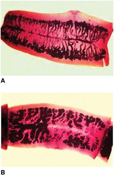
The species of the proglottid can be identified by pressing the segment between two glass microscope slides and counting the main lateral branches of the uterus. T solium usually has 7 to 13 branches on each side; T saginata usually has 15 to 20 lateral branches on each side (Fig. 340-1). Fecal examination, especially with T saginata infection, often is unrewarding because intact gravid proglottids tend to be eliminated or crawl out onto the perianal area before they disintegrate and release their eggs. Thus, the perianal cellophane-tape method, similar to that used to diagnose pinworms, may be more effective for recovering Taenia ova.
Approximately 10% of patients with neurocysticercosis have eosinophilia. The findings on lumbar puncture are rarely helpful, and findings range from normal to isolated high protein levels with or without an inflammatory pleocytosis. Eosinophilia may be present occasionally in the CSF. A lumbar puncture should not be done in the presence of suspected increased intracranial pressure.
Radiographic findings are often useful. Soft-tissue radiographic studies may reveal characteristic numerous, tiny, curvilinear calcifications in the muscle. MRI or CT will demonstrate cysts in all stages in the meninges and parenchyma (Fig. 340-2). Contrast-enhancement studies with metrizamide often are necessary to demonstrate isodense cysts in the ventricles.
In the past, ELISA has been the most frequently used diagnostic method to detect cysticercus antibodies in both serum and cerebrospinal fluid (CSF). This test can be highly sensitive, but may cross-react with other helminth antibodies, especially Echinococcus. The enzyme-linked immunoelectrotransfer blot (EITB) is highly specific and sensitive, although sensitivity is low when fewer than 2 parenchymal cysts are present. In a recent series of children presenting with neurocysticercosis in the United States, fewer than 30% had positive EITB. Examination of the serum is more sensitive than the CSF. In patients with clinical and radiologic features of cysticercosis, negative serology may be an indication for biopsy, especially if the patient is from an area of low endemicity. Elevated titers in CSF are particularly useful if they exceed those in the serum. High positive titers are more often seen in those individuals with hydrocephalus or meningeal involvement.
 TREATMENT
Adult tapeworm infections are treated successfully if the scolex is eliminated. An effective agent with few untoward effects is niclosamide (Yomesan) but this agent is only inconsistently available in the United States. For Taenia infections, the single dose for adults consists of 4 tablets or 2 g chewed thoroughly after a light meal. For children weighing 11 to 34 kg, a single dose of 2 tablets (1 g) is recommended, and for those children weighing more than 34 kg, a single dose of 3 tablets (1.5 g) is recommended. For patients with T solium infection, therapy probably should be administered in the physician’s office. An antiemetic may be administered 30 minutes before the antihelminthic. If the patient does not have a bowel movement within 2 hours, a mild saline purge should be provided. Alternatively, praziquantel, an acylated isoquinole-pyrazine, is highly active against most tapeworm infections. It can be given in a single dose of 10 to 20 mg/kg in taeniasis.

Stay updated, free articles. Join our Telegram channel

Full access? Get Clinical Tree


