Suprasellar Mass
Susan I. Blaser, MD, FRCPC
DIFFERENTIAL DIAGNOSIS
Common
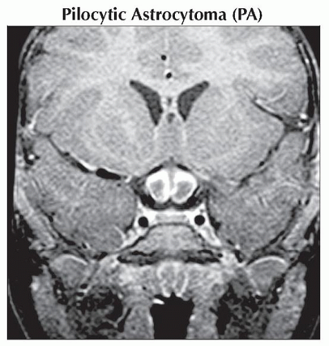
Pilocytic Astrocytoma (PA)
Craniopharyngioma
Pituitary Hyperplasia (Physiologic)
Hydrocephalus
Less Common
Germinoma
Tuber Cinereum Hamartoma
Arachnoid Cyst
Langerhans Cell Histiocytosis
Pituitary Stalk Anomalies
Teratoma
Rare but Important
Lipoma
Pituitary Macroadenoma
Dermoid Cyst
Leukemia
Pilomyxoid Astrocytoma
Saccular Aneurysm
Retinoblastoma (Trilateral)
Lymphocytic Hypophysitis
Lymphoma, Primary CNS
Rathke Cleft Cyst
ESSENTIAL INFORMATION
Key Differential Diagnosis Issues
Is mass extra- or intraaxial?
Extraaxial masses arise from pituitary/infundibulum, meninges, vessels
If extraaxial mass appears to arise from pituitary/infundibulum, determine origin of mass as precisely as possible
Pituitary gland: Think physiologic hyperplasia, hypophysitis, macroadenoma (rare in children)
Infundibular stalk: Germinoma, histiocytosis; stalk anomalies, lymphoma, leukemia (rare)
Nonpituitary extraaxial masses (normal pituitary gland can usually be identified inferior to lesion)
Craniopharyngioma
Hydrocephalus
Arachnoid cyst
Saccular aneurysm
Intraaxial masses arise from chiasm/hypothalamus/3rd ventricle
Optic chiasm/hypothalamus: Pilocytic or pilomyxoid astrocytoma, tuber cinereum hamartoma, lipoma
Third ventricle: Hydrocephalus > > neoplasm
T1 hyperintense suprasellar mass in child? Think craniopharyngioma, lipoma, dermoid, posterior pituitary ectopia
Helpful Clues for Common Diagnoses
Pilocytic Astrocytoma (PA)
Most occur in children 5-15 years old
Enlarged optic nerve/chiasm/tract
Usually solid, iso-/hypointense on T1WI; hyperintense on T2WI, FLAIR
Variable enhancement (none to intense)
If large, bulky H-shaped mass in infant, may be pilomyxoid variant
Craniopharyngioma
90% Ca++ (globular, rim)
90% cystic (may have multiple)
90% enhance (rim, nodule)
Density/signal intensity within cysts/locules varies with content
Pituitary Hyperplasia (Physiologic)
Up to 10 mm height, convex superior margin in young menstruating females
“Macroadenoma-appearing” mass in child?
May be hyperplasia, not tumor (especially prepubescent male)!
Hydrocephalus
Enlarged 3rd ventricle (aqueductal stenosis, obstructive hydrocephalus)
Anterior recesses protrude inferiorly
May enlarge bony sella over time
Helpful Clues for Less Common Diagnoses
Germinoma
50-60% involve pituitary gland/stalk
Often presents with diabetes insipidus (DI)
Tuber Cinereum Hamartoma
Isosexual precocious puberty > gelastic seizures
Pedunculated (“collar button”) or sessile mass between infundibular stalk, mamillary bodies
Can be tiny (1-2 mm) or giant (3-5 cm)
Isointense with gray matter (occasionally slightly hyperintense on FLAIR)
Doesn’t enhance
Arachnoid Cyst
10% suprasellar
Sharply marginated CSF-like cyst
Sagittal T1- or T2WI shows 3rd ventricle elevated, compressed over cyst
Suppresses on FLAIR, DWI negative
Langerhans Cell Histiocytosis
Child usually < 2 years old
May have central DI
10% of LCH cases involve stalk, pituitary gland ± hypothalamus
Rare: Choroid plexus, leptomeninges, cerebellar WM, brain parenchyma
Look for solitary/multiple lytic skull lesions with “beveled edges”
Pituitary Stalk Anomalies
Posterior pituitary ectopia
Short stature ± endocrine deficiencies
Posterior pituitary “bright spot” missing
Mislocated along tuber cinereum
Stalk small/absent
Duplicated pituitary gland/stalk
Endocrinologically normal
± midline facial anomalies
Tuber cinereum/mamillary bodies fused
Teratoma
Optic chiasm > pineal
Ca++, cysts, soft tissue, fat
Helpful Clues for Rare Diagnoses
Lipoma
Fatty hypothalamic mass
Pituitary Macroadenoma
“Figure of 8” pituitary mass
Gland cannot be separated from mass
Dermoid Cyst
Fat-like mass ± droplets in CSF
Fat suppression sequences confirm
20% Ca++
Leukemia
Rare; look for other lesions (sinuses, dura)
Pilomyxoid Astrocytoma
Rare variant of PA
Large, bulky suprasellar mass in infant
May hemorrhage (rare in PA)
Saccular Aneurysm
Rare in children (< 2% of all saccular aneurysms occur in pediatric age group)
When occur, often large/bizarre
Thrombus common
Look for residual patent lumen, phase artifact
Retinoblastoma (Trilateral)
3rd tumor in pineal or suprasellar region
Lymphocytic Hypophysitis
Adolescent > child
May cause DI
Can mimic macroadenoma, pituitary apoplexy
Lymphoma, Primary CNS
Rare in children
Can mimic hypophysitis, germinoma, LCH
Rathke Cleft Cyst
Rare in children
Cyst in/above pituitary, separate from stalk
Rarely calcifies, does not enhance (“claw” of enhancing pituitary tissue may surround mass)
Intracystic nodule virtually pathognomonic
Image Gallery
 Coronal T1 C+ MR shows chiasmatic glioma. The prechiasmatic optic nerves are expanded and surrounded by enhancing tumor. |
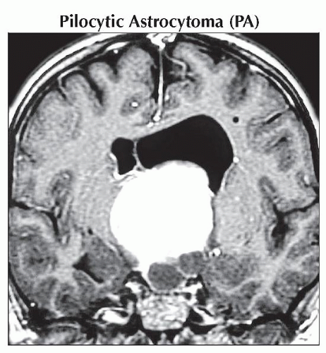 Coronal T1WI C+ MR shows a very large suprasellar pilocytic astrocytoma. This solid and cystic mass involves the suprasellar cistern, the chiasm, the hypothalamus and protrudes into the 3rd ventricle. |
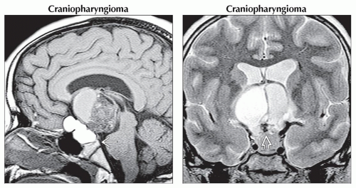 (Left) Sagittal T1WI MR shows typical cysts of varying signal intensity in the suprasellar cistern, herniating into the 3rd ventricle. There is enlargement of the bony sella and erosion of the dorsum sella
 . (Right) Coronal T2WI MR shows calcification . (Right) Coronal T2WI MR shows calcification  at the base of the lesion. at the base of the lesion.Stay updated, free articles. Join our Telegram channel
Full access? Get Clinical Tree
 Get Clinical Tree app for offline access

|