Objective
Enhanced myometrial vascularity (EMV) is a clinically significant risk factor that puts women at risk for severe hemorrhage, which is often difficult to control. Such bleeding can result in the need for blood transfusion, uterine artery embolization (UAE), or even hysterectomy. Recognizing high-risk EMV early can help detect patients who are at risk for bleeding. Although the peak systolic velocity (PSV) is used to determine the severity of EMV and its associated risk of complications, to date there is no standardized method for evaluation and diagnosis of in patients with suspected EMV.
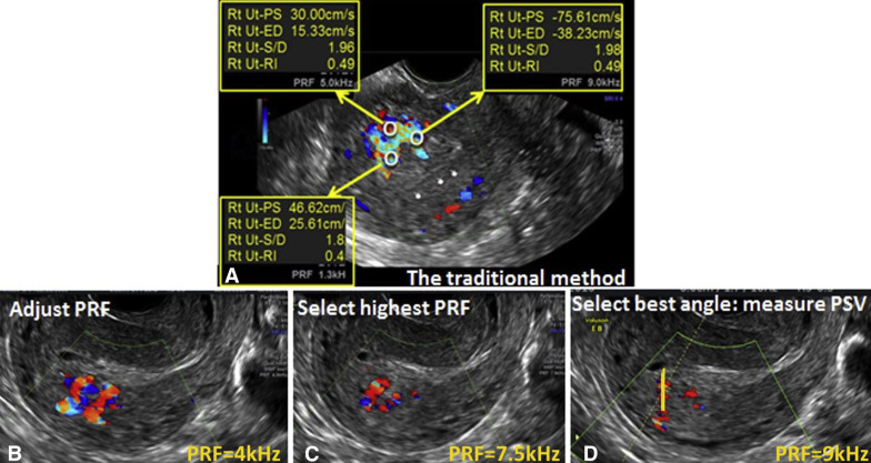
This study aimed to propose a general standard for the measurement of PSV in EMV by comparing the 2 different protocols ( Figure 1 ).

Study Design
In this institutional review board–approved study, EMV was evaluated in 14 patients with the use of 2 separate protocols. The first, what we termed the traditional protocol , included measuring PSV at several subjectively chosen vessels and selecting the highest PSV value. The second, our defined standardized protocol , involved 3 steps: First, a “panoramic” gray scale image of the uterus was obtained, followed by spectral pulse wave Doppler image that focused on suspicious areas. Second, pulse repetition frequency (PRF) was increased gradually until only a few vessels were seen to isolate the vessels with the highest blood flow velocity. Finally, the highest PSV was measured by angle correction.
We compared the PSV value measured using the traditional protocol with the PSV obtained using the standardized protocol. PSV of ≥83 cm/s was classified as high risk for bleeding based on historical studies, which used the traditional protocol. PSV of the 2 protocols were compared with the use of the Wilcoxon nonparametric test.
Stay updated, free articles. Join our Telegram channel

Full access? Get Clinical Tree


