Fig. 38.1
CT scan showing enlarged uterus with cystic spaces and thinned out myometrium
A differential diagnosis of uterine sarcoma/endometrial malignancy was made on imaging. CA125 and LDH were within normal limits. Ultrasound guided Trucut biopsy was taken and reported as smooth muscle neoplasm with no significant mitosis on histopathology. Peroperatively uterus was large, soft to firm in consistency with normal tubes and ovaries. Total hysterectomy with bilateral salphingo-oopherectomy was done. The specimen was sent for frozen section and reported as smooth muscle neoplasm with degenerative changes. Malignancy was excluded.
Final histopathology report with immunohistochemistry was consistent with STUMP.
Macroscopic description: Uterus 26 × 19 cm. Sectioning showed an intramural mass of 18 × 13 cms. Endometrium was mildly thickened.
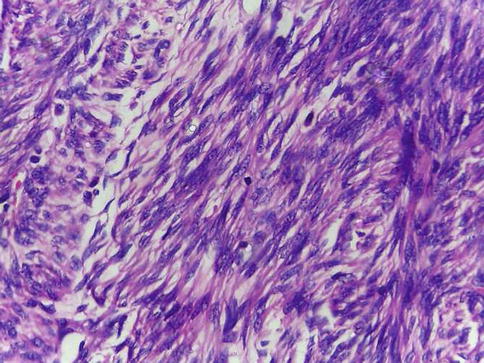
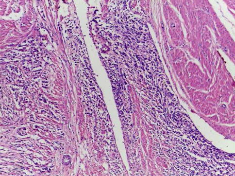
Microscopy: Spindle cell neoplasm with occasional mitosis without pleomorphism or necrosis, with infiltrating margins (Figs. 38.2 and 38.3)


Fig. 38.2
Cellular tumor area with a mitotic figures

Fig. 38.3
Infiltrative margins
Immunohistochemistry: KI 67showed no proliferative activity.
P53: Occasional cells showed positivity.
Bcl2 was positive.
Chest CT was done postoperatively and was within normal limits. Patient is on close follow up and 12 months following surgery she has no evidence of recurrent disease.
References
1.
2.
Kempson RL. Sarcomas and related neoplasms. In: Noris HJ, Hertig AT, Abel MR, editors. The uterus. Baltimore: Williams and Wilkins; 1973.
3.
Bell SW, Kempson RL, Hendrickeson MR. Problematic uterine smooth muscleneoplasms: a clinicopathological study of 213cases. Am J Surg Pathol. 1994;18:535–58.CrossRefPubMed
Stay updated, free articles. Join our Telegram channel

Full access? Get Clinical Tree


