Renal Morphogenesis
Sunder Sims-Lucas and Carlton M. Bates
The process of renal morphogenesis provides a basis for understanding the causes of kidney aplasia, dysplasia, and hypoplasia, which together are the second leading cause of chronic renal insufficiency in children. It also may provide insight into congenital nephron underendowment, which may not present as overt renal disease in childhood but has been linked with adult-onset diseases such as hypertension and end-stage renal failure.1,2
OVERVIEW OF RENAL MORPHOGENESIS
The permanent mammalian kidney or meta-nephros forms following the development of transient excretory precursors, the pronephros and the mesonephros. The pronephros, the first urinary system in vertebrates, appears at 22 days of human embryogenesis in the cervical region (Fig. 464-1).3 Each pronephric nephron consists of a simple glomus that filters the blood through tubular epithelial cords called nephrotomes.4 While the pronephros is not functional in mammals and eventually regresses, it must form or there will be complete renal (metanephric) agenesis.5 The mesonephros, the second urinary system in vertebrates, appears at about the fourth week of human gestation, immediately caudal to the pronephric ducts (Fig. 464-1).3 Unlike the pronephros, the mesonephros is functional in mammals during embryogenesis. While most of the mesonephros ultimately regresses, portions of the mesonephric duct in males form the vas deferens, the seminal vesicle, and part of the epididymis, while some mesonephric tubules persist as testicular efferent ductules. In females, the mesonephros usually degenerates completely due to the presence of müllerian inhibitory substance.
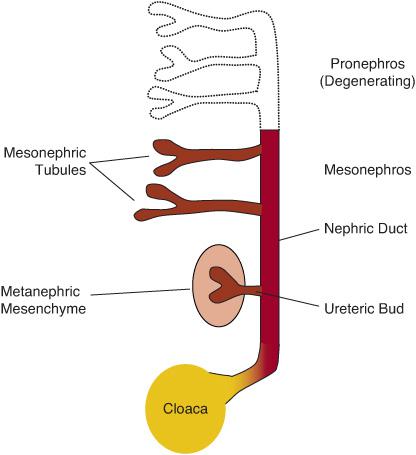
FIGURE 464-1. Mammalian embryonic kidney development. Mammalian kidney development proceeds in a craniocaudal direction beginning with formation of the pronephros, then the mesonephros, and finally the metanephros, or the adult kidney. Although transitory, the pronephros and mesonephros are necessary for development of the metanephros. The metanephros begins when the ureteric bud invades the metanephric mesenchyme.
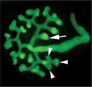
The metanephros, the definitive kidney in mammals, first appears in the fifth week of human gestation in the region of the hind-limb (Fig. 464-1).3 Paired densities of mesenchyme, termed metanephric mesenchyme, secrete molecules that induce an epithelial outgrowth from the mesonephric duct called the ureteric bud, which then invades the mesenchyme. Once these 2 tissues make contact, they begin a series of reciprocal signaling events leading to formation of the meta-nephric kidney. The mesenchymal cells induce the ureteric duct to elongate and branch fairly dichotomously (Fig. 464-2) to ultimately form the collecting ducts, pelvis, and ureter. Signals from the ureteric bud tips in turn stimulate the mesenchymal cells to condense and then convert into nephron epithelia (Fig. 464-3). Surrounding the developing nephrons and ureteric bud tree is stromal mesenchyme, which develops into the interstitial tissues of the adult kidney. There is also an extensive vascular network that develops in the metanephric kidney.
DEVELOPMENT OF THE METANEPHRIC KIDNEY
Much of the genetic programming of the pronephros and mesonephros mimic that of the metanephros, and absence of the mesonephros leads to metanephric agenesis. Each step of metanephric formation and the molecular signals regulating these steps are outlined in the following sections. Most of these molecules have been characterized in mice, given the ability to manipulate genes globally or in specific tissues such as the kidney.6-11
 URETERIC BUD OUTGROWTH
Ureteric bud outgrowth from the mesonephric (wolffian) duct is one of the earliest stages of metanephric kidney development. The bud initially appears as an evagination from the wolffian duct that then elongates and makes direct contact with the surrounding metanephric mesenchyme. Under normal circumstances, there is only 1 ureteric bud induction site per wolffian duct. The metanephric mesenchyme secretes glial cell line–derived neurotrophic factor (GDNF) signals that bind to RET (a receptor tyrosine kinase) and GDNF family receptor alpha-1 (GFRα1, a coreceptor), both of which are present in the wolffian duct.93-98 Recent studies have also begun to identify human renal abnormalities associated with GDNF/RET mutations.101,102
Stay updated, free articles. Join our Telegram channel

Full access? Get Clinical Tree


