Fig. 30.1
Four-year-old child with femur fracture treated by traction in a Thomas’ splint. (a) Injury film. (b) X-ray in Thomas’s splint. (c) At 2 months fracture united with 40° angulation and 3 cm shortening. The malunion is due to muscle forces that were not effectively managed
Deformities following trauma to the extremities are not uncommon and not always dependent on the level of care provided. While some deformities are inevitable due to high-energy trauma and damage to the growing area of long bones, other deformities occur because of suboptimal management allowing the limb to be positioned abnormally or allowing certain muscles to get tight and create a secondary deformity.
Deformities that are not clinically significant or have the potential for substantial remodeling to an acceptable level can be observed. In general, when the residual axial malalignment of a lower extremity long bone exceeds 10°, correction may be warranted.
Causation and Etiological Factors
Box 30.1. Factors Related to Development of Posttraumatic Deformities
Injury related
Soft tissue
Skin and soft-tissue loss
Tendon and muscle injury
Nerve injury
Vascular injury
Skeletal
Type of fracture, fracture geometry, and comminution
Growth plate injury
Bone loss
Intra-articular injury
Treatment related
Missed diagnosis of a fracture
Delayed presentation and management
Incorrect treatment choice
Suboptimal management
Overtreatment
Iatrogenic problems
Complication related
Infection
Loss of reduction
Compartment syndrome
Postoperative stiffness
Avascular necrosis
Patient-related factors
Preexisting skeletal problems like dysplasia, syndromes
Neuromuscular problems like cerebral palsy, poliomyelitis, muscular dystrophy
Altered bone physiology due to osteogenesis imperfecta, metabolic bone disease
Pathological fracture with underlying tumor, osteomyelitis
Immunocompromise
Malnutrition
A posttraumatic deformity can be a result of initial soft-tissue and skeletal injury, its management, or complications. In order to accurately identify the cause and anticipate the natural progression of the deformity, it is important to ascertain the mechanism of injury, initial soft-tissue and bony injury, specifics of the treatment given—conservative and operative, postoperative care, and complications and course of the deformity thus far.
Box 30.2. Etiological Mechanisms for Posttraumatic Deformities
Postural or positional
Muscle forces
Neural and vascular injury
Poor skeletal stabilization
Damage to the growing area of bone
Infection
Deformities can be caused by one or more of the following etiological factors:
1.
Postural or positional: Lack of immobilization or faulty immobilization may cause a deformity, often related to contracture of the adjacent soft tissues. Examples are an equinus deformity of the foot or a knee flexion deformity because of contracture of the gastro-soleus or the hamstrings, respectively.
2.
Muscle forces: Varus deformity of the upper femur following a fracture is a classic example of muscle forces at work. The flexor-adductor predominance over the abductor-extensor forces causes this deformity (e.g., Figs. 30.1 and 30.2).
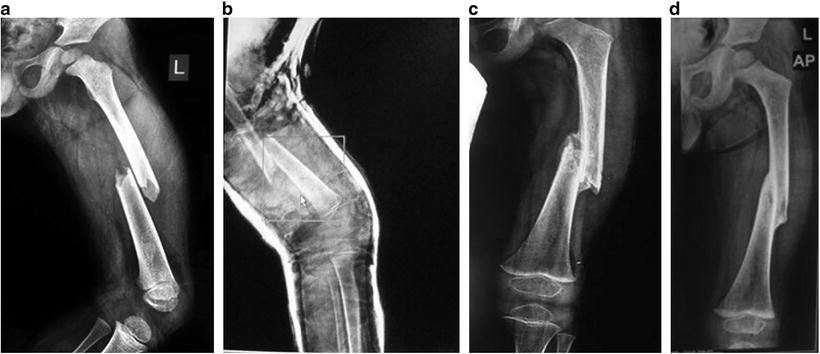
Fig. 30.2
Effect of remodeling and growth: Fracture shaft femur in a 4-year-old child. (a) Injury film. (b) Position in plaster after immediate spica showing shortening and recurvatum. (c) At 6 weeks, uniting with 20 varus and 2.5 cm shortening. (d) At 3 months, united with persistent shortening. (e) After 2 years, correction of deformity with equal limb lengths
3.
Neural or vascular injury: Neural injury can create deformity because of imbalance of muscle forces or by muscle paralysis causing a postural deformity. Vascular injury may give rise to infarction of muscle followed by its fibrosis and eventually a contracture. Volkmann’s ischaemic contracture is a typical example of such an etiology.
4.
Poor skeletal stabilization, loss of reduction, and loss of fixation can all give rise to posttraumatic deformities. Factors such as poor bone quality, traumatic bone loss, and fracture comminution may contribute to inadequate skeletal stabilization (e.g., see Fig. 30.9).
5.
Damage to the growing area of bone: Classic examples of these are partial physeal injuries with growth arrest giving rise to various angular deformities (e.g., see Fig. 30.7). Symmetric physeal damage would give rise to limb shortening. Damage to the growing area may be a consequence of the mode of injury or may occur because of its poor management. Salter–Harris type 6 injury is a perichondrial ring injury presenting with a normal X-ray. This is an example of the mode of injury giving rise to deformity (e.g., see Fig. 30.10).
6.
Infection: Open skeletal injuries or iatrogenic issues can be complicated by infection which works by damaging bone and cartilage, causing bone lysis and loss, growth plate damage and its sequelae, as well as joint ankylosis and deformity.
Classification
Posttraumatic deformities can be classified into different ways to fully understand the current problems as well as potential issues that may arise in the future. Such classification helps in formulating the management plan for the individual patient.
Box 30.3. Classification of Posttraumatic Deformities in Children
Related to the area of bone affected
Diaphyseal
Metaphyseal
Physeal
Epiphyseal
Related to the anatomical effect
Angulation
Rotation
Limb length
Nonunion
Restriction of movement
Muscle weakness
Related to the tissue affected
Soft tissue
Skin, fascia, muscle, capsule, or ligaments
Skeletal
Diaphyseal, growth plate, articular
Related to the progression
Progressive: Improving: Partial or complete
Progressive: Improving, then progressive
Clinical Manifestations
Fixed deformity of the joint, joint stiffness and ankylosis, shortening, angular and torsional deformities, and axial malalignment are all potential clinical manifestations in a posttraumatic situation. Meticulous clinical assessment may point to the static or progressive nature of the deformity. Presence or absence of infection is to be noted as it is an important consideration in management. Examination for neural injury is important and can be challenging in young children, especially in the presence of fixed contractures.
Investigations
Besides plain X-rays, MRI and CT scans may be useful in some patients.
Plain X–rays are the simplest way of monitoring bony deformity. In addition to standard views one may need X-rays in the plane of the deformity. Full-length standing radiographs and scanograms are useful for monitoring limb length.
MRI can give a lot of information about the growth plate (e.g., see Fig. 30.7f, g) and articular cartilage, periarticular ligamentous injury, osteochondral fractures, and articular alignment.
CT scans help define the fracture geometry, area of growth arrest, and presence of a bony bar. It is also a useful tool to quantify torsional deformities.
Occasionally, specialized scans like the radioisotope scans may be required to detect infection. Hematologic tests including a white cell count, sedimentation rate, and C-reactive protein are helpful in such a situation. A more comprehensive metabolic panel may be required in patients with underlying metabolic bone diseases such as various forms of rickets.
Remodeling of Bony Deformities
Remodeling may impact decision making for correcting any bone deformity. The patient’s functional capacity and the surgeon’s experience should also be factors in determining whether to depend on the remodeling capacity of the specific fracture or to consider performing a more aggressive, invasive technique to achieve a satisfactory result [1].
In the typical long bone, 75 % of the remodeling occurs by reorientation of the physis while appositional remodeling of the diaphysis can only be expected to contribute 25 % to the remodeling process.
Important factors influencing remodeling are as follows:
1.
Age of the patient: The older the child the less is the remodeling potential. In general, remodeling of posttraumatic deformities occurs more effectively in children younger than 10 years.
2.
Location of the malunion: Metaphyseal fractures remodel better than diaphyseal fractures. Fractures near the fast-growing physes such as lower femur and upper tibia and lower radius and upper humerus remodel better than their counterparts.
3.
The plane of the deformity: Angulation in the plane of the joint axis corrects better. Rotations do not correct for all practical purposes. In some fractures overgrowth phenomenon gives rise to correction of limb shortening.
4.
Magnitude of angulation and translation.
Box 30.4. Factors Affecting Remodeling of Fractures in Children
Age
Location
Plane of deformity
Degree of deformity
Some guidelines on acceptability of fracture reduction and remodeling are given at the end of this chapter (see section Acceptability Criteria for Reduction in Lower Limb Fractures).
Case Study (Fig. 30.2)
Clinical Summary
A 4-year-old child was treated for fracture shaft femur by immediate spica. He had 2-cm shortening and varus deformity 3 months post-fracture. Over a period of 2 years, the deformity corrected and child had equal leg lengths.
Philosophy of Treatment
Fracture healing can stimulate bone growth in certain femoral shaft fractures. The amount of overgrowth varies in different reports from 0.4 to 2.7 cm. Overgrowth in femoral fractures appears to be independent of age, fracture level, and position of the fracture at the time of healing. The effect of growth stimulation may continue for up to 3 years following the fracture.
Case Study (Fig. 30.3)
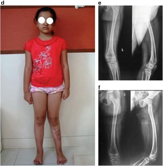
Fig. 30.3
A 6-year-old with malunited type 1 SH fracture. (a) Four months from injury, Feb 2011. (b) MRI showing posterior displacement of the physis. (c, d) X-ray and clinical picture 5 months postinjury. (e) X-rays 10 months postinjury showing remodeling and correction of angulation and translation. (f) Complete remodeling at 1.5 years postinjury in 2012
Clinical Summary
A 6-year-old girl presented 4 months after an open fracture of the tibial tuberosity with patellar tendon tear which was debrided, repaired, and cast. She developed a stiff knee for which the original surgeon did a manipulation under anaesthesia. However, the local swelling and deformity increased. A Salter–Harris type 1 injury was diagnosed at presentation. An MRI revealed a posterior displacement of the distal femoral epiphysis with loculated fluid collection along the posterior border.
Different options of management were considered but it was decided to leave her alone in view of the good possibility of remodeling. For the stiff knee physical therapy with range of motion exercises was started. The sequential X-rays show the excellent remodeling (Table 30.1). She regained nearly full knee movement 2 years after the injury.
Table 30.1
Diaphyseal-epiphyseal angle
Feb 2011 | 38° |
Mar 2011 | 42° |
May 2011 | 54° |
Aug 2011 | 70° |
Nov 2011 | 82° |
Apr 2012 | 90° |
Philosophy of Management
The distal femoral epiphysis is one of the fastest growing physes with rapid remodeling in children. Undisplaced Salter–Harris type 1 injuries need not be fixed, as long as they are immobilized with restricted weight bearing and closely followed with serial radiographs. The ones with displacement may need a gentle reduction and fixation. We prefer that pins not be introduced from the knee level upwards but rather in a reverse fashion from the metaphysis into the epiphysis as recommended by Wall [2]. It is important not to leave pins sticking out of the skin if placed retrogradely. If placed in this fashion they should be buried under the skin for later removal.
Management
Decision making in posttraumatic deformity situations is based on a number of factors, including:
Age of the patient
Location of the deformity
Morphological status of bone, soft tissue, and cartilage
Functional loss and impairment
Abnormal mechanical loading with risk of joint degeneration
Cosmetic aspect
Table 30.2 presents a general schema of the types of interventions.
Table 30.2
General schema of the types of interventions
Type of deformity | Nature of intervention |
|---|---|
Joint contractures | Manipulation |
Splintage | |
Serial plasters | |
Soft-tissue release | |
External fixation | |
Osteotomy | |
Arthrodesis | |
Muscle imbalance | Splintage |
Tendon transfer | |
Arthrodesis | |
Angular and rotational bony deformities | Osteotomy |
Growth modulation | |
Physeal bar excision | |
Shortening | Shoe raise |
Limb lengthening | |
Contralateral epiphysiodesis | |
Limb shortening | |
Bone loss and nonunion | Bone grafting |
Bone transport |
Important Posttraumatic Deformities: Lower Limb
In this section we briefly discuss some unique posttraumatic lower limb deformities in children and demonstrate a variety of treatment strategies. However, as noted earlier, the decision making should be individualized based on patient, surgeon, and environmental factors.
Posttraumatic Chondrolysis of the Hip and Avascular Necrosis
Acetabular injuries may be complicated by chondrolysis of the hip, and this may give rise to a fixed flexion and adduction deformity. Likewise hip dislocations and fractures of the femoral neck may be complicated by avascular necrosis. Avascular necrosis may follow the commonly described patterns in the classifications of Kalamchi and MacEwen [3] or of Bucholz and Ogden [4] and management would follow the lines described for such conditions.
Stay updated, free articles. Join our Telegram channel

Full access? Get Clinical Tree


