Fig. 60.1
a and b Clinical photographs showing a very large ovarian teratoma causing marked abdominal distension
Teratomas are usually benign tumors . They have a characteristic appearance and teeth, bone, and hair are found inside the tumor.
They are subdivided into mature teratomas, which are benign, or immature teratomas which may be either malignant or benign (Figs. 60.2 and 60.3).
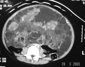
Fig. 60.2
CT scan of the abdomen and pelvis showing a huge mass occupying most of the abdomen
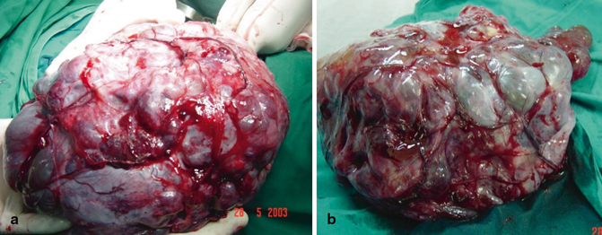
Fig. 60.3
a and b Intraoperative photographs showing a large ovarian tumor which turned out to be an immature ovarian teratoma
Most benign teratomas are composed of mature cells, but 20–30 % also contain immature elements, most often neuroepithelium.
The tumors may be picked up on plain film due to the presence of calcification in two-thirds of teratomas (Fig. 60.4).
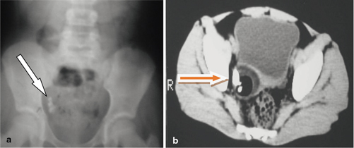
Fig. 60.4
Abdominal x-ray (a) and CT scan (b) showing pelvic calcification in an ovarian teratoma
Malignant Germ Cell Tumor
These tumors include:
1.
Yolk sac tumors
2.
Choriocarcinoma
3.
Immature tearatomas
Benign Cystic Teratomas (Dermoid Cysts)
This is the most common benign ovarian tumor in childhood and is composed of mature, well-differentiated tissue.
Approximately 10 % are bilateral (Fig. 60.5).
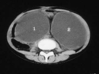
Fig. 60.5
CT scan showing bilateral ovarian cysts
About 50 % will have a calcification visible on x-ray.
The average age of patients with benign ovarian teratomas is 12 years.
These teratomas tend to undergo torsion.
Dermoid cysts are normally treated with oophorectomy.
Dysgerminoma
The second most common ovarian tumor in children after the teratoma.
It is the most common malignant ovarian tumor in children and adolescents.
The tumor is typically low-grade and on imaging it is solid, smooth, and well encapsulated.
Of the dysgerminomas, 20 % are bilateral.
Usually grow to a large size before diagnosis.
Dysgerminomas are usually nonfunctioning tumors.
These tumors can spread locally and are known to be very radiosensitive.
They have a good prognosis with > 90 % survival.
Rhabdomyosarcoma
It is the most common soft tissue sarcoma in children and a frequent cause of death.
Roughly 10 % of all solid tumors in childhood are Rhabdomyosarcomas.
These tumors are extremely malignant and tend to invade locally and metastases by hematogenous and through lymphatic routes.
Without treatment, 90 % of patients die within 1 year of diagnosis.
Rhabdomyosarcomas occur equally in males and females and are seen in two age peaks at 4–6 years and 16–18 years of age.
The different histologic types that can be seen in a rhabdomyosarcoma include alveolar, embryonal, embryonal-botryoid, pleomorphic, and undifferentiated.
Among these types, the alveolar variety has the worst prognosis.
Stay updated, free articles. Join our Telegram channel

Full access? Get Clinical Tree


