Ovarian Cancer


Ovarian Cancer


Pedro T. Ramirez

David M. Gershenson


INCIDENCE AND RISK FACTORS

The incidence of ovarian cancer is highest in Sweden (19.6/100,000) and the United States (15.4/100,000) and lowest in Japan (10.1/100,000). In the United States, ovarian cancer incidence rates are highest in Caucasian women, intermediate in African American women, and lowest in Native American women.

Factors associated with an increase in ovarian cancer risk are age, nulliparity, and a family history of the disease. Ovarian cancer is rare before the age of 40, increases steadily thereafter, and peaks at ages 65 to 75. Parity is the most important nongenetic factor affecting risk for ovarian cancer (Table 53.1). Whittimore and colleagues analyzed 12 case-control studies in the United States and reported a significant risk reduction for ovarian cancer with each term pregnancy (odds ratio [OR] = 0.47). The risk of ovarian cancer decreased progressively with increasing numbers of pregnancies. Similarly, the use of oral contraceptives has been shown to reduce the risk of ovarian cancer. Ovarian cancer risk decreases approximately 11% per year of oral contraceptive use to a maximum of 46% after 5 years of use. These observations have led to the theory of “incessant ovulation” in the etiology of ovarian cancer. According to this theory, risk for epithelial ovarian cancer is related directly to the number of uninterrupted ovulatory cycles. With ovulation, the surface epithelium is ruptured and then undergoes rapid proliferation and repair. At the time of ovulation, there is invagination of the surface epithelium into the underlying stroma forming inclusion cysts. The epithelium lining these inclusion cysts then undergoes neoplastic transformation under the influence of oncogenic factors. The observation that early age of menarche and late menopause are associated with an increase in ovarian cancer risk is consistent with this theory, because both increase the number of ovulatory cycles.

A second theory of ovarian carcinogenesis is the retrograde menstruation hypothesis. According to this hypothesis, retrograde transportation of carcinogens from the uterus and lower genital tract through the fallopian tube to the ovary occurs at the time of menstruation. The protective effect of oral contraceptives is consistent with this hypothesis because their use has been associated with a reduction in menstrual blood loss, thus with decreased retrograde menstruation. Conversely, the observed increase in ovarian cancer risk associated with hormone replacement therapy (Table 53.1) may be mediated through the periods of abnormal uterine bleeding that occur with many hormonal regimens. This hypothesis is also supported by the known decrease in ovarian cancer risk in women who have undergone tubal ligation or hysterectomy, because these procedures prevent the ascent of potential oncogenic factors from the lower genital tract to the ovary.


TABLE 53.1 Risk Factors for Ovarian Cancer


FACTOR


ODDS RATIO


Paritya


Nulliparous


1


Parous


0.47


Oral contraceptive useb


Never


1


2-5 y


0.73


Estrogen useb


Never


1c


Ever


1.4


Clomid used


No


1


Yes


2.3


Tubal ligationa


No


1


Yes


0.59


Hysterectomya


No


1


Yes


0.66


Talc usee


No


1


Yes


1.5


Breast-feedinga


No


1


Yes


0.73


Family historyf


1 first-degree relative with ovarian cancer


3.1


>2 first-degree relatives with ovarian cancer


4.6


a Whittemore AS, Harris R, Itnyre J. Collaborative Ovarian Cancer Group: characteristics relating to ovarian cancer risk, collaborative analysis of 12 U.S. case-control studies. II. Invasive epithelial ovarian cancers in white women. Am J Epidemiol 1992;136:1184.

b Riman T, Dickman PW, Nilsson S, et al. Hormone replacement therapy and the risk of invasive epithelial ovarian cancer in Swedish women. J Natl Cancer Inst 2002;94:497.

c Not significant.

d Rossing MA, Daling JR, Weiss NS, et al. Ovarian tumors in a cohort of infertile women. N Engl J Med 1994;331:771.

e Cook LS, Kamb ML, Weiss NS. Perineal powder exposure and risk of ovarian cancer. Am J Epidemiol 1997;145:459.

f Kerlikowske K, Brown JS, Grady DG. Should women with familial ovarian cancer undergo prophylactic oophorectomy? Obstet Gynecol 1992;80:700.


A final hypothesis concerning the genesis of ovarian cancer is that exposure of ovarian epithelium to persistently high levels of pituitary gonadotropins results in neoplastic transformation. Follicle-stimulating hormone (FSH) has been shown to promote the growth of epithelial ovarian cancer cells in vitro,
and this effect can be blocked by luteinizing hormone (LH). A corollary to this hypothesis is that elevated circulating gonadotropin levels promote estrogen biosynthesis in the ovarian stroma, which, in turn, causes abnormal proliferation of the adjacent epithelium. Breast-feeding, which has been reported to lower the risk of ovarian cancer, is associated with reduced serum concentrations of LH and estradiol. Pregnancy and the use of oral contraceptives presumably lower the risk of ovarian cancer by inhibiting pituitary secretion of gonadotropins. This theory also receives support from the observed increased risk of ovarian cancer in women taking fertility drugs because these drugs stimulate ovulation by increasing levels of FSH, particularly in the follicular phase of the cycle.

Perhaps the most important risk factor for epithelial ovarian cancer is a family history of the disease. The estimated odds ratio for the development of ovarian cancer in a woman with one first-degree relative who has ovarian cancer is 3.1 (95% CI = 2.1 to 4.5). This risk increases even further (OR 4.6 CI = 1.1 to 18.4) in a woman with two or more primary or secondary relatives who have ovarian cancer. These odds ratios translate to a lifetime risk for ovarian cancer of approximately 5% in a woman with one first-degree relative who has the disease and 7% in a woman with two or more relatives with the disease. It should be mentioned, however, that familial ovarian cancers make up a relatively small proportion of total ovarian cancer cases. Only 5% to 10% of ovarian cancer patients report having a positive family history of the disease.

Three familial ovarian cancer syndromes have been described: Hereditary breast-ovarian cancer syndrome (HBOC), hereditary site-specific ovarian cancer syndrome (HSSOC), and hereditary nonpolyposis colon cancer syndrome (HNPCC). HBOC, the most common of the familial syndromes, is characterized by multiple cases of early-onset (<50 years of age) breast and ovarian cancers. This syndrome accounts for 75% to 90% of all hereditary ovarian cancer cases. HSSOC is manifested only by an increase in cases of early-onset ovarian cancer and makes up about 5% of hereditary ovarian cancers. Women with HSSOC are often younger and more commonly have tumors with serous histology than do women with sporadic ovarian cancer. HNPCC, or Lynch syndrome type II, is characterized by a predominance of early-onset proximal colon cancer in association with cancers of the endometrium and ovary. HNPCC is often confirmed by a mutation in one of several mismatch repair genes, in particular MLH1, MSH2, and MSH6. The estimated lifetime risk of ovarian cancer in women with HNPCC is 10% to 12%. These three familial ovarian cancer syndromes are inherited by autosomal dominant transmission through either maternal or paternal lineage. Therefore, the children of an affected parent have a 50% risk of inheriting the genetic abnormality.

Germline mutations in the BRCA1 or BRCA2 gene appear to account for most hereditary ovarian cancers. BRCA1 was identified in 1994 and is located on the long (q) arm of chromosome 17. BRCA1 is thought to be a tumor suppressor because the normal copy of BRCA1 is always deleted in ovarian cancers that arise in women who inherit a mutant BRCA1 gene. It is estimated that germline mutations in BRCA1 are responsible for 80% to 90% of hereditary ovarian cancers. BRCA2 was identified in 1995 and is located on the long (q) arm of chromosome 13. In a recent report from the Breast Cancer Linkage Consortium, BRCA1 mutations were identified in 81% of ovarian cancer families, whereas BRCA2 mutations were detected in 14% of those families. Penetrance is variable (range 10% to 50%) from one individual to another, and it is estimated that the lifetime risk of ovarian cancer is approximately 39% in BRCA1 carriers and 11% in BRCA2 carriers.


SIGNS AND SYMPTOMS

Although most reports indicate that patients with early-stage ovarian cancer have few symptoms, a recent national survey of 1,725 ovarian cancer patients provides evidence that many of these patients actually had symptoms that they or their primary health providers ignored. The most common symptoms of patients with stage I or II ovarian cancer were abdominal bloating or pain, indigestion, urinary frequency, and constipation. Because many of these symptoms are nonspecific, patients were unaware that they could be associated with ovarian cancer. As a result, 22% of patients ignored their symptoms entirely, and 30% reported that the wrong diagnosis was made. A pelvic examination was performed in only two thirds of patients, and 45% had a delay in diagnosis of more than 3 months. Patients with advanced disease commonly reported abdominal swelling, fatigue, and weight loss. In a subsequent study, four target symptoms were significantly more common in ovarian cancer patients before diagnosis than in age-matched control patients. These symptoms included abdominal pain (frequency 30%, OR 6), abdominal swelling (frequency 16.5%, OR 30), gastrointestinal (GI) symptoms (frequency 8.5%, OR 2.3), and pelvic pain (frequency 5.4%, OR 4.3). These observations emphasize the need for patient and physician education concerning the possible relationship of rather nonspecific abdominal symptoms to ovarian cancer. A high index of suspicion, coupled with pelvic ultrasound and CA-125 testing, may lead to the earlier diagnosis of ovarian cancer in selected patients with these symptoms. Routine yearly pelvic examination has not been shown to increase the diagnosis of early-stage ovarian cancer.

Although vaginal bleeding is not commonly associated with ovarian cancer, it may be present in patients with metastatic involvement of the uterus. Likewise, endometrial hyperplasia and abnormal uterine bleeding can be caused by excess estrogen production from an ovarian stromal tumor. Ovarian cancer must be considered in a patient who presents with a pelvic mass and shortness of breath. A malignant pleural effusion from metastatic ovarian cancer is more common on the right side and is usually associated with dullness to percussion and decreased breath sounds. Finally, any patient with a clinically detected pelvic tumor on pelvic examination should undergo careful palpation of both groins to rule out inguinal lymphadenopathy secondary to metastatic disease.


PREOPERATIVE EVALUATION

Before operative intervention, each patient should undergo a thorough evaluation designed to determine the anatomic location, size, and morphology of the ovarian tumor, as well as possible sites of metastases. In addition, her general medical condition and ability to undergo a major surgical procedure should be established. All patients should undergo routine hematologic and biochemical testing. A computed tomography (CT) scan provides valuable information concerning cardiac size, as well as the presence of pulmonary metastases or a pleural effusion. Positron emission tomography (PET) scans may be more sensitive, but their value in the preoperative assessment of primary ovarian cancer has not been proven.

CT scanning with contrast may identify ureteral obstruction, retroperitoneal lymphadenopathy, omental disease, and peritoneal metastases. In a patient with ascites but no ovarian tumor, liver function studies should be performed to exclude cirrhosis or liver disease. Rarely, the presence of right heart failure and hepatic congestion will cause ascites. Paracentesis may be useful in a patient who presents with ascites and
no evidence of an ovarian abnormality. The characteristics of malignant cells present in ascitic fluid may help identify the primary site of intra-abdominal malignancy. An electrocardiogram is indicated in all women older than age 40 or in a patient with specific signs or symptoms of cardiac disease.


TABLE 53.2 Serum Markers in Ovarian Cancer


TUMOR HISTOLOGY


SERUM MARKER


Epithelial ovarian cancer


CA-125


Mucinous cystadenocarcinoma


CEA


Endodermal sinus tumor


AFP


Embryonal cell carcinoma


hCG, AFP


Choriocarcinoma


hCG


Dysgerminoma


LDH-1, LDH-2


Granulosa cell tumor


Inhibin


AFP, alpha-fetoprotein; CEA, carcinoembryonic antigen; hCG, human chorionic gonadotropin; LDH, lactate dehydrogenase.


Common sites of nongynecologic cancer that spreads to the ovary include gastric malignancy, colonic carcinoma, and breast carcinoma. A careful history and physical examinations should arouse suspicion of other possible primary malignancies. Any such possibilities should be evaluated with appropriate ongoing studies and diagnostic evaluation, possibly including colonoscopy, upper GI endoscopy, and/or mammography. A biopsy of a suspicious finding or easily accessible lymph node may clarify the diagnosis.

Finally, serum markers should be obtained according to the age and clinical findings of each patient. Serum CA-125 and CEA often are elevated in patients with epithelial ovarian cancer, whereas serum alpha-fetoprotein, human chorionic gonadotropin, or lactate dehydrogenase is more commonly increased in younger women with germ-cell ovarian malignancies. Serum inhibin is the most reliable marker in patients with ovarian granulosa cell tumors. The specific marker associated with each type of ovarian cancer is illustrated in Table 53.2. It is important to obtain a baseline serum marker value before surgery so that it can be used to monitor response to therapy.


PATTERNS OF SPREAD

Ovarian cancer spreads by (a) direct extension and exfoliation of tumor cells into the peritoneal cavity, (b) lymphatic metastases to regional and paraaortic lymph nodes, and (c) hematogenous dissemination (Fig. 53.1). The specific pattern of spread depends on the stage, cell type, and histologic differentiation of the tumor.

The earliest method of spread in epithelial ovarian cancer is by exfoliation of tumor cells from the ovarian surface. These cells migrate with the circulation of peritoneal fluid along the surfaces of the pelvic and mesenteric peritoneum. They also are carried cephalad in the paracolic spaces to the omentum and undersurface of the diaphragm. Spread to the right lung occurs through the transdiaphragmatic lymphatics in the right hemidiaphragm, often producing a right pleural effusion. Surface spread to the peritoneal surfaces of the bowel and bladder is a common finding in advanced-stage ovarian cancer, but involvement of the bowel lumen or bladder mucosa is rare.


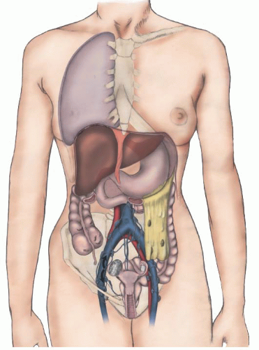
FIGURE 53.1 Patterns of spread of ovarian cancer.

Lymphatic drainage from the ovary follows two pathways. The first involves lateral spread through the broad ligament to the pelvic lymph nodes. In patients with advanced-stage disease, there may be retrograde dissemination via the lymphatics of this pathway to the round ligament to the inguinal lymph nodes. The second pathway of efferent lymphatic drainage follows the ovarian vein to the paracaval and paraaortic lymph nodes. Metastatic spread of ovarian cancer to lymph nodes is well documented even in early-stage disease (Table 53.3) and confirms that there may be separate pathways of dissemination to the pelvic and paraaortic lymph nodes. Cass and coworkers, for example, reported that 14 of 96 patients (15%) with disease visibly confined to one ovary had microscopic lymph node metastases. All 14 patients with nodal spread had poorly differentiated tumors. Isolated ipsilateral lymph node metastases occurred in five patients, and isolated contralateral lymph node metastases occurred in three patients. Pelvic lymph nodes were involved in six patients, paraaortic lymph nodes in five patients, and both in three patients. As expected, the frequency of lymph node metastases is related to stage of disease, cell type, and histologic differentiation of the tumor. Chen and Lee, for example, reported that the frequency of pelvic lymph node metastases increased from 9% in patients with clinically apparent stage I ovarian cancer to 33% in patients with stage IV disease. Similarly, the frequency of paraaortic lymph node involvement increased from 18% in patients with clinically apparent stage I disease to 67% in patients with stage IV disease. These findings are similar to those of Burghardt and colleagues, who noted lymph node metastases in 74% of patients
with stage III or IV ovarian cancer. The incidence of lymph node metastases increased from 20% in well-differentiated ovarian cancers to 65% in poorly differentiated tumors and was higher in serous ovarian malignancies than in mucinous or endometrioid cancers.


TABLE 53.3 Lymph Nodal Metastases in Patients with Clinically Apparent Stage I Epithelial Ovarian Cancer


AUTHOR


PATIENTS


PELVIC LYMPH NODE METASTASES


PARAAORTIC LYMPH NODE METASTASES


Onda et al. (1996)


30


6 (18%)


5 (15%)


Carnino et al. (1997)


47


1 (4%)


0 (0%)


Burghardt et al. (1991)


20


3 (15%)


1 (5%)


Sakai et al. (1997)


46


1 (2%)


1 (2%)


Chen and Lee (1983)


11


1 (9%)


2 (18%)


Li et al. (2000)


91


9 (10%)


8 (9%)


Total


245


21 (9%)


17 (7%)


Hematogenous spread of ovarian cancer to the parenchyma of the liver or lung is fortunately quite rare (<5%) at the time of initial diagnosis, but may occur, particularly in poorly differentiated tumors that become refractory to combination chemotherapy.


EXAMINATION OF OVARIAN TUMOR SPECIMENS AND HISTOLOGIC CLASSIFICATION

Ovarian tumor specimens should be described, fixed, and sectioned according to the guidelines established by the College of American Pathologists (CAP). Tumors should be classified histologically according to the World Health Organization classification and nomenclature of ovarian tumors (Table 53.4).
Although there are numerous grading systems for ovarian cancers that use both architectural and nuclear features, it is recommended that four grades be used, with grade 4 (undifferentiated) applied to tumors with minimal or no differentiation. Recommendations concerning the use of special staining techniques or flow cytometry in establishing the correct histologic diagnosis of ovarian tumors are made in the CAP report. Immunohistochemical staining of ovarian tumors for cytokeratin 7 (CK7) and cytokeratin 20 (CK20) is helpful in differentiating primary mucinous ovarian carcinoma from colorectal adenocarcinoma that has metastasized to the ovary. Colorectal adenocarcinomas usually stain positively for CK20 and negatively for CK7. In contrast, ovarian carcinomas usually stain negatively for CK20 and positively for CK7.


TABLE 53.4 World Health Organization Histological Classification of Ovarian Tumors


Surface epithelial-stromal tumors


Serous tumors


Malignant


Adenocarcinoma


Surface papillary adenocarcinoma


Adenocarcinofibroma (malignant adenofibroma)


Borderline tumor


Papillary cystic tumor


Surface papillary tumor


Adenofibroma, cystadenofibroma


Benign


Cystadenoma


Surface papilloma


Adenofibroma and cystadenofibroma


Mucinous tumors


Malignant


Adenocarcinoma


Adenocarcinofibroma (malignant adenofibroma)


Borderline tumor


Intestinal type


Endocervical-like


Benign


Cystadenoma


Adenofibroma and cystadenofibroma


Mucinous cystic tumor with mural nodules


Mucinous cystic tumor with pseudomyxoma peritonei


Endometrioid tumors, including variants with squamous differentiation


Malignant


Adenocarcinoma, not otherwise specified


Adenocarcinofibroma (malignant adenofibroma)


Malignant mullerian mixed tumor (carcinosarcoma)


Adenosarcoma


Endometrioid stromal sarcoma (low grade)


Undifferentiated ovarian sarcoma


Borderline tumor


Cystic tumor


Adenofibroma and cystadenofibroma


Benign


Cystadenoma


Adenofibroma and cystadenofibroma


Clear cell tumors


Malignant


Adenocarcinoma


Adenocarcinofibroma (malignant adenofibroma)


Borderline tumor


Cystic tumor


Adenofibroma and cystadenofibroma


Benign


Cystadenofibroma


Adenofibroma and cystadenofibroma


Transitional cell tumors


Malignant


Transitional cell tumor (non-Brenner type)


Malignant Brenner tumor


Borderline


Brenner tumor


Proliferating variant


Benign


Brenner tumor


Metaplastic variant


Squamous cell tumors


Squamous cell carcinoma


Epidermoid cyst


Mixed epithelial tumors (specify components)


Malignant


Borderline


Benign


Undifferentiated and unclassified tumors


Undifferentiated carcinoma


Adenocarcinoma, not otherwise specified


Sex cord-stromal tumors


Granulosa-stromal cell tumors


Granulosa cell tumor group


Adult granulosa cell tumor


Juvenile granulosa cell tumor


Thecoma-fibroma group


Thecoma, not otherwise specified


Typical


Luteinized


Fibroma


Cellular fibroma


Fibrosarcoma


Stromal tumor with mixed sex cord elements


Sclerosing stromal tumor


Signet-ring stromal tumor


Unclassified (fibrothecoma)


Sertoli-stromal cell tumors


Sertoli-Leydig cell tumor group (androblastomas)


Well differentiated


Of intermediate differentiation


Variant with heterologous elements (specify type)


Poorly differentiated (sarcomatoid)


Variant with heterologous elements (specify type)


Retiform


Variant with heterologous elements (specify type)


Sertoli cell tumor


Stromal-Leydig cell tumor


Sex cord-stromal tumors of mixed or unclassified cell types


Sex cord tumor with annular tubules


Gynandroblastoma (specify components)


Sex cord-stromal tumor, unclassified


Steroid cell tumors


Stromal luteoma


Leydig cell tumor group


Hilus cell tumor


Leydig cell tumor, nonhilar type


Leydig cell tumor, not otherwise specified


Steroid cell tumor, not otherwise specified


Well differentiated


Malignant


Germ-cell tumors


Primitive germ-cell tumors


Dysgerminoma


Yolk sac tumor


Polyvesicular vitelline tumor


Glandular variant


Hepatoid variant


Embryonal carcinoma


Polyembryoma


Nongestational choriocarcinoma


Mixed germ-cell tumor (specify components)


Biphasic or triphasic teratoma


Immature teratoma


Mature teratoma


Solid


Cystic


Dermoid cyst


Fetiform teratoma (homunculus)


Monodermal teratoma and somatic-type tumors associated with dermoid cysts


Thyroid tumor group


Struma ovarii


Benign


Malignant (specify type)


Carcinoid group


Insular


Trabecular


Mucinous


Strumal carcinoid


Mixed


Neuroectodermal tumor group


Ependymoma


Primitive neuroectodermal tumor


Medulloepithelioma


Glioblastoma multiforme


Others


Carcinoma group


Squamous cell carcinoma


Adenocarcinoma


Others


Melanocytic group


Malignant melanoma


Melanocytic nevus


Sarcoma group (specify type)


Sebaceous tumor group


Sebaceous adenoma


Sebaceous carcinoma


Primary-type tumor group


Retinal anlage tumor group


Others


Germ-cell sex cord-stromal tumors


Gonadoblastoma


Variant with malignant germ-cell tumor


Mixed germ-cell/sex cord-stromal tumor


Variant with malignant germ-cell tumor


Tumors of the rete ovarii


Adenocarcinoma


Adenoma


Cystadenoma


Cystadenofibroma


Miscellaneous tumors


Small-cell carcinoma, hypercalcemic type


Small-cell carcinoma, pulmonary type


Large-cell neuroendocrine carcinoma


Hepatoid carcinoma


Primary ovarian mesothelioma


Wilms tumor


Gestational choriocarcinoma


Hydatidiform mole


Adenoid cystic carcinoma


Basal cell tumor


Ovarian wolffian tumor


Paraganglioma


Myxoma


Soft tissue tumors not specific to the ovary


Others


Tumorlike conditions


Luteoma of pregnancy


Stromal hyperthecosis


Stromal hyperplasia


Fibromatosis


Massive ovarian edema


Others


Lymphoid and hematopoietic tumors


Malignant lymphoma (specify type)


Leukemia (specify type)


Plasmacytoma


Secondary tumors


PRIMARY SURGERY


Patterns of Care in Ovarian Cancer Surgery

All patients with suspected ovarian cancer should be referred to a gynecologic oncologist for evaluation and possible surgery. Unfortunately, in both Europe and the United States (where most of the outcomes studies have been conducted), too high a proportion of women with ovarian cancer are receiving substandard care. The role of comprehensive staging, particularly pelvic and paraaortic lymphadenectomy, in patients with early-stage ovarian cancer has been a topic of wide discussion, primarily because many women with earlystage disease are inadequately staged and because surgeons with the expertise to perform a lymphadenectomy are not always involved with the initial surgery. A study by Goff et al. (2006) that evaluated patterns of surgical care across the United States revealed that among women with earlystage disease, 21.4% underwent an oophorectomy with or without a hysterectomy but without any additional staging or debulking procedure and 47% did not have nodal sampling. Carney and associates found that less than 50% of ovarian cancer patients in Utah were cared for by a gynecologic oncologist. In addition, they also noted that care by a gynecologic oncologist was associated with a survival advantage. In the Maryland study, Bristow and coworkers found that the majority of ovarian cancer patients in Maryland were undergoing primary surgery in low-volume settings.

In patients with early-stage disease, lymphadenectomy, in addition to providing prognostic information, may have therapeutic value. Chan et al. evaluated 6,686 women diagnosed with clinical stage I ovarian cancer to estimate the survival impact of lymphadenectomy. The authors found that lymphadenectomy was associated with improved 5-year disease-specific survival. In addition, the extent of lymphadenectomy was significantly associated with the 5-year disease-specific survival rate (0 nodes sampled, 87%; fewer than 10 nodes sampled, 92%; and 10 or more nodes sampled, 94%; P < 0.001). In a study of the SEER program database from 1991 and 1996, Harlan and colleagues found that comparing the two treatment periods, the majority of women with early-stage ovarian cancer did not receive guideline therapy in both periods, although the rate was improved in the 1996 cohort. For women with advanced-stage ovarian cancer, there was a higher rate of guideline therapy use but no improvement between 1991 and 1996—62.6% versus 62.3%. The totality of this information clearly underscores the need for improvement in patterns of referral.


Early-Stage Ovarian Cancer


Comprehensive Surgical Staging


STEPS IN THE PROCEDURE


Comprehensive Staging of Early Disease (Laparoscopy)


  • Entry into abdominal cavity and inspection of pelvic and peritoneal cavity


  • Cytology obtained


  • Affected ovary isolated by identification of infundibulopelvic ligament


  • Ureter identified and isolated


  • Infundibulopelvic ligament cauterized and transected


  • Utero-ovarian ligament cauterized and transected


  • Ovary placed in endoscopic bag intact and removed through largest trocar site assuring no spilling of ovarian tissue


  • Frozen section obtained and if consistent with cancer, then formal staging performed


  • Omental biopsy performed by coagulation and transection of dessicated tissue


  • Pelvic and paraaortic lymph node dissection is performed bilaterally.


  • Multiple peritoneal biopsies performed including both paracolic gutters, pelvic peritoneal surfaces bilaterally, and diaphragm peritoneum


Ovarian cancer is staged according to the International Federation of Gynecology and Obstetrics (FIGO) staging system (Table 53.5). The official FIGO staging for ovarian cancer was extensively changed in 2012 and has been recently published. Staging is based on a thorough surgical evaluation of the possible metastatic spread of the cancer and histologic evaluation of the primary ovarian lesion. It is too early to know if substaging of stage IC or IIIA will be reliable prognostic characteristics in general, nonresearch settings. Should preoperative evaluation suggest an area of extra-abdominal or intrahepatic metastasis, fine needle aspiration or needle biopsy of this lesion should be performed. Because ovarian cancer frequently spreads to upper abdominal structures, a vertical midline abdominal incision is recommended. This incision should be extended high enough to remove the primary ovarian tumor and to evaluate the stomach, omentum, liver, and undersurface of the diaphragm. Rupture of a cystic ovarian malignancy is associated with a poorer prognosis, so the incision should be sufficient to allow excision of the primary tumor with its capsule intact. Once the abdomen has been opened, the following steps should be performed for adequate surgical staging (Table 53.6):


  • The volume of ascitic fluid should be recorded, and a minimum of 25 mL should be sent for cytologic evaluation.


  • In the absence of ascites, separate saline washings should be obtained from the (a) pelvic cul-de-sac, (b) right paracolic space, (c) left paracolic space, and (d) undersurface of each hemidiaphragm. Approximately 100 mL of saline should be instilled in each of these areas, recovered, and sent for cytologic evaluation.


    TABLE 53.5 FIGO Ovarian Cancer Staging (January 01, 2014)


    STAGE I: Tumor confined to ovaries


    OLD


    NEW


    IA


    Tumor limited to 1 ovary, capsule intact, no tumor on surface, negative washings/ascites


    IA


    Tumor limited to 1 ovary, capsule intact, no tumor on surface, negative washings


    IB


    Tumor involves both ovaries


    IB


    Tumor involves both ovaries otherwise like IA


    IC


    Tumor involves 1 or both ovaries with any of the following: capsule rupture, tumor on surface, positive washings/ascites


    IC Tumor limited to 1 or both ovaries


    IC1


    Surgical spill


    IC2


    Capsule rupture before surgery or tumor on ovarian surface


    IC3


    Malignant cells in the ascites or peritoneal washings


    STAGE II: Tumor involves 1 or both ovaries with pelvic extension (below the pelvic brim) or primary peritoneal cancer


    OLD


    NEW


    IIA


    Extension and/or implant on uterus and/or fallopian tubes


    IIA


    Extension and/or implant on uterus and/or fallopian tubes


    IIB


    Extension to other pelvic intraperitoneal tissues


    IIB


    Extension to other pelvic intraperitoneal tissues


    IICa


    IIA or IIB with positive washings/ascites


    STAGE III: Tumor involves 1 or both ovaries with cytologically or histologically confirmed spread to the peritoneum outside the pelvis and/or metastasis to the retroperitoneal lymph nodes


    OLD


    NEW


    IIIA


    Microscopic metastasis beyond the pelvis


    IIIA (Positive retroperitoneal lymph nodes and /or microscopic metastasis beyond the pelvis)


    IIIA1


    Positive retroperitoneal lymph nodes only


    IIIA1(i) Metastasis ≤ 10 mm


    IIIA1(ii) Metastasis > 10 mm


    IIIA2


    Microscopic, extrapelvic (above the brim) peritoneal involvement ± positive retroperitoneal lymph nodes


    IIIB


    Macroscopic, extrapelvic, peritoneal metastasis ≤2 cm in greatest dimension


    IIIB


    Macroscopic, extrapelvic, peritoneal metastasis ≤2 cm ± positive retroperitoneal lymph nodes. Includes extension to capsule of liver/spleen


    IIIC


    Macroscopic, extrapelvic, peritoneal metastasis >2 cm in greatest dimension and/or regional lymph node metastasis


    IIIC


    Macroscopic, extrapelvic, peritoneal metastasis >2 cm ± positive retroperitoneal lymph nodes. Includes extension to capsule of liver/spleen


    STAGE IV: Distant metastasis excluding peritoneal metastasis


    OLD


    NEW


    IV


    Distant metastasis excluding peritoneal metastasis. Includes hepatic parenchymal metastasis.


    IVA


    Pleural effusion with positive cytology


    IVB


    Hepatic and/or splenic parenchymal metastasis, metastasis to extra-abdominal organs (including inguinal lymph nodes and lymph nodes outside of the abdominal cavity)


    a Old stage IIC has been eliminated.


    Other major recommendations are as follows:


    • Histologic type including grading should be designated at staging.


    • Primary site (ovary, fallopian tube, or peritoneum) should be designated where possible.


    • Tumors that may otherwise qualify for stage I but involved with dense adhesions justify upgrading to stage II if tumor cells are histologically proven to be present in the adhesions.


    Reprinted from Prat J, for the FIGO Committee on Gynecologic Oncology. Staging classification for cancer of the ovary, fallopian tube and peritoneum. Intl J Gynecol Obstet 2014;124:1-5. Copyright © 2013 International Federation of Gynecology and Obstetrics. Published by Elsevier Ireland Ltd. All rights reserved. Reprinted with permission from Elsevier.


    TABLE 53.6 Surgical Staging of Apparent Early-Stage Ovarian Cancer


    Vertical midline incision


    Evacuation of ascites or multiple cytologic washings


    Complete abdominal inspection and palpation


    Resection of ovaries, fallopian tubes, and uterusa


    Omentectomy


    Random peritoneal biopsies


    Retroperitoneal lymph node sampling


    a Exceptions may be made in selected patients who wish to preserve fertility.


  • The ovarian tumor should be inspected, with particular attention to the presence of papillary excrescences on the surface or rupture of the capsule. The contralateral ovary and uterus should be examined for the presence of metastatic tumor. The pathways of ovarian tumor should be removed and sent for frozen section examination. Removal of the opposite ovary and/or uterus is dependent on several factors and is discussed on the following pages.


  • Careful inspection and palpation of the peritoneal surfaces and intra-abdominal viscera should be performed. This evaluation should be approached in a systematic fashion, beginning with the peritoneum of the cul-de-sac and small bowel mesentery. Inspection should continue with the ascending colon, liver, omentum, undersurface of the right and left hemidiaphragms, and stomach. Finally, the transverse colon, spleen, descending colon, and bladder peritoneum should be evaluated.


  • All areas suspicious for malignancy should be biopsied. In the absence of visible disease, biopsies should be taken of the cul-de-sac peritoneum, bladder peritoneum, both lateral pelvic walls, paracolic peritoneum bilaterally, and undersurface of the right hemidiaphragm. An infracolic omentectomy should be performed in patients with epithelial ovarian cancer and an omental wedge biopsy taken in patients with germ-cell or stromal tumors. Appendectomy should be performed in all patients with mucinous epithelial cancers involving the ovary. Primary appendiceal cancers, although rare, commonly spread to the ovaries and usually require right hemicolectomy as part of initial surgical staging.


  • As has been mentioned, ovarian cancer commonly spreads to both pelvic and paraaortic lymph nodes. Some patients with early-stage ovarian cancer have paraaortic lymph node metastases in the absence of pelvic lymph node spread. Therefore, these lymph node groups should be sampled separately in all patients. It is important that sampling include lymph nodes on the opposite side of the primary ovarian tumor, because isolated contralateral spread has been reported. In the setting of advanced-stage disease, prospective randomized trials reviewed by Panici et al. have shown that routine lymphadenectomy is not associated with a higher survival rate; however, it is associated with an improvement in the disease-free interval.


  • Finally, it should be emphasized that operative findings present at the time of staging must be carefully documented. Prognosis is related to the site and volume of metastatic tumor, as well as the amount of residual disease remaining after surgical debulking. Important data concerning the location and size of tumor metastases are often lost if the details concerning operative staging are not recorded.


TABLE 53.7 Criteria for Potential Fertility-Sparing Surgery in Ovarian Cancer Patients


Patient desirous of preserving fertility


Patient and family consent and agree to close follow-up


No evidence of dysgenetic gonads


Specific situations


Any unilateral malignant germ-cell tumor


Any unilateral sex cord-stromal tumor


Any unilateral borderline tumor


Stage IA invasive epithelial tumor


Fertility-Sparing Surgery

Although the majority of ovarian malignancies occur in older women for whom bilateral salpingo-oophorectomy and hysterectomy are standard treatment, a significant subset of patients is young and can be managed more conservatively (Table 53.7). Conservative management is used here to denote surgery that preserves reproductive potential without compromising curability. With some exceptions, such a strategy may be applicable for women younger than 40 years who wish to bear children.

When contemplating surgery on a young patient with a suspected ovarian malignancy, it is important to discuss with her all possible operative findings and procedures and the longterm implications of the various options. If the patient is a child, the parents need to clearly understand this information. In most instances, young patients have their initial surgery done outside major university hospitals or cancer centers. Common errors in surgical management include incomplete surgical staging and unnecessary bilateral salpingo-oophorectomy. In addition, some patients are mismanaged because of an error in the pathologic diagnosis of a rare ovarian neoplasm.

The optimal candidate for conservative surgical management is a young patient who has stage IA disease. If, on initial inspection, the suspected cancer is confined to one ovary, then unilateral salpingo-oophorectomy is appropriate. If the mass is thought to be benign, then ovarian cystectomy may be preferable. The specimen should be sent for frozen section examination. If malignancy is diagnosed, then appropriate staging biopsies should be performed, as discussed. If the contralateral ovary appears normal, it is recommended that it not be biopsied to avoid potential infertility caused by peritoneal adhesions or ovarian failure.

One should not rely too heavily on frozen section examination in making the decision to perform a hysterectomy and bilateral salpingo-oophorectomy. If the histologic diagnosis is in question, it is always preferable to wait for permanent section results for a young patient, even if this requires a repeat surgery.

The advent of in vitro fertilization technology has been a significant impact on intraoperative management. Previously, convention dictated that if a bilateral salpingo-oophorectomy is necessary, a hysterectomy should also be performed. However, current technology for donor oocyte transfer and hormonal support allows a woman without ovaries to sustain an intrauterine pregnancy. Similarly, if the uterus and one ovary are resected because of tumor involvement, current techniques allow retrieval of oocytes from the patient’s remaining ovary, in vitro fertilization with sperm from her male partner, and implantation of the embryo into a surrogate’s uterus. Therefore, traditional guidelines concerning surgical management of ovarian cancer may no longer be applicable in selected young patients.


Approximately 50% to 70% of malignant germ-cell tumors are stage I. Except for dysgerminoma, in which the incidence of bilaterality is 10% to 15%, bilateral ovarian tumors are exceedingly rare. Such a finding almost always signifies advanced disease with metastatic spread from one ovary to the other or a mixed germ-cell tumor with a dysgerminoma component. Benign cystic teratoma is associated with malignant germ-cell tumors in 5% to 10% of cases and may occur in one or both ovaries. Therefore, unilateral salpingo-oophorectomypreserving the contralateral ovary and uteruscombined with surgical staging can be performed in most patients with malignant germ-cell tumors, even many with advancedstage disease. If the contralateral ovary is enlarged, most likely it represents a benign cystic teratoma that can be managed with an ovarian cystectomy only. With the exception of stage IA pure dysgerminoma or stage IA, grade 1 immature teratoma, patients with germ-cell tumors require postoperative chemotherapy consisting of the combination of bleomycin, etoposide, and cisplatin (BEP). Several series have documented normal reproductive function in at least 80% of patients as well as pregnancies following fertility-sparing surgery and chemotherapy.

In the past few years, some investigators have advocated the practice of surgery alone for other categories of malignant germ-cell tumors, including stage IA, grade 2 and 3 immature teratomas, and stage IA yolk sac tumors. Such an approach, however, should be taken with caution; many experts would still consider it experimental. Nevertheless, there is a trend toward careful observation following surgery alone for most patients with stage I malignant ovarian germ-cell tumors. The Children’s Oncology Group is currently conducting a clinical trial that includes postoperative surveillance for patients with stage I disease. Chemotherapy is initiated only for serum tumor marker elevation.

Most sex cord-stromal tumors are confined to the ovary. Stage I accounts for greater than 50% (in some series as high as 100%) of granulosa cell tumors. More than 90% of Sertoli-Leydig cell tumors are stage IA. Bilaterality occurs in less than 5% of cases with either tumor type. Therefore, optimal surgical management of most patients with stromal tumors consists of unilateral adnexectomy. There does not appear to be a role for lymphadenectomy in patients with sex cord-stromal tumors. A study by Brown et al. evaluated 262 patients with sex cord-stromal tumors. Of these, 111 patients underwent a complete or partial staging procedure. None of the patients had evidence of positive nodes. The authors also noted that of the 117 patients whose disease eventually recurred, 6 patients (5.1%) had nodal metastases at the time of recurrence. Endometrial biopsy or curettage also should be performed in any young patient whose uterus is preserved because 5% to 15% of patients with granulosa cell tumors develop endometrial cancer or hyperplasia. Postoperative therapy may be indicated for patients with metastatic disease or for selected patients with stage I disease (e.g., poorly differentiated Sertoli-Leydig cell tumor or granulosa cell tumor with rupture). The most commonly used regimens in this setting include either BEP or the combination of paclitaxel and carboplatin.

Approximately 10% to 15% of all ovarian neoplasms are of the borderline or low malignant potential classification. Although they were first described more than 70 years ago, only in the last few years have we begun to fully appreciate their biologic behavior. Approximately 33% to 60% of serous borderline tumors are limited to one ovary. Extraovarian spread is noted in approximately 20% to 30% of cases. Approximately 80% to 90% of mucinous borderline tumors are confined to one ovary. Both endometrioid and clear cell borderline tumors are almost always stage I, and the vast majority is unilateral. For patients with borderline tumors seemingly confined to one ovary, appropriate surgical management includes unilateral salpingo-oophorectomy with surgical staging. The use of ovarian cystectomy instead of unilateral adnexectomy also has been reported, although some patients treated in this manner require repeat surgery for a recurrence of tumor in the same or opposite ovary. If bilateral borderline tumors are present, portions of one or both ovaries may be preserved with ovarian cystectomy, if feasible. Whatever the surgical approach, reported 5-year survival rates for patients with stage I borderline tumors treated with surgery alone are 95% or better.

For patients with borderline tumors and peritoneal implants, the optimal management remains controversial. However, surgical excision is the mainstay of treatment. After frozen section confirmation of borderline tumor, an effort should be made to resect all gross disease. In addition, staging biopsies of peritoneal surfaces and lymph nodes are indicated, as are cytologic washings. However, there is no consensus regarding the extent of surgical staging required, particularly with regard to the need for lymphadenectomy. A recent study was conducted to evaluate the prognostic value of lymph node involvement for borderline tumors. This was a retrospective study on 49 patients at a single institution, and a broader analysis was also performed on 1,503 patients obtained from the Surveillance, Epidemiology and End Results (SEER) database. The authors concluded that lymph node involvement does not appear as a prognosis factor in patients with advanced-stage borderline tumors. Even in the face of metastatic disease, a normal contralateral ovary may be preserved in young patients. In patients with advanced-stage serous disease, the incidence of bilateral tumors is approximately 75%. There are several reports detailing successful pregnancies following treatment in patients with borderline ovarian tumors.

The relapse risk for patients with peritoneal implants is related to the type: noninvasive versus invasive. For patients with noninvasive peritoneal implants, the lifetime risk is estimated to be at least 20%. For those with invasive peritoneal implants, the lifetime risk of relapse is estimated to be at least 50%. Consequently, postoperative platinum-based chemotherapy is recommended for the latter diagnosis by many groups. However, no study has demonstrated a survival benefit related to postoperative chemotherapy.

Invasive epithelial tumors account for approximately 70% of all ovarian malignancies. Despite the low overall survival rate associated with these tumors, selected young patients with stage I disease can be treated conservatively. The major factors that influence the selection process are histologic grade and bilaterality, in addition to stage. Serous tumors are bilateral in about 50% of cases. The incidence of bilaterality for mucinous tumors varies widely in reported series from as low as 5% to as high as 50%, but probably is no greater than 10% to 20%. Approximately 30% to 50% of endometrioid and clear cell cancers are bilateral.

Only gold members can continue reading. Log In or Register to continue

Stay updated, free articles. Join our Telegram channel

Jun 4, 2016 | Posted by in GYNECOLOGY | Comments Off on Ovarian Cancer

Full access? Get Clinical Tree

Get Clinical Tree app for offline access