8.1 Orthopaedic problems
See Chapter 12.2 for Bone and joint infections.
Skeletal variations during growth
Parents can be anxious about whether their child is normal. They refer to adult posture, which is different to that of infants and children. These ‘developmental’ postures can be due to:
• intrauterine posture, sometimes described as ‘packaging’
• developmental variants – not present at birth, but may appear during growth and then disappear spontaneously. These include the common conditions of bow legs, knock knees, flat feet and in-toeing. These conditions seldom require active treatment but parents do need informed reassurance, which must be based on accurate knowledge of the natural history of these variations of posture in infants and children.
Intrauterine posture
The position of the child before birth is normally one of flexion. The spine is flexed so that it forms a long curve with a concavity forward, the arms and legs are flexed, and the feet may assume a variety of postures. In the newborn the intrauterine posture can be readily reconstructed by ‘folding’ the baby into his or her most comfortable position, and this may indicate any postural abnormality present.
Two common foot postures are seen in newborns.
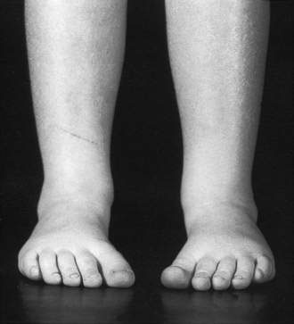
Talipes calcaneovalgus
Many babies are born with the foot turned upwards at the ankle so that the toes lie close to the front of the shin: this is known as talipes calcaneovalgus (Fig. 8.1.1). This posture can be corrected passively so that the foot can be brought down to a plantigrade position or even into equinus. The condition has a strong tendency to correct itself spontaneously over a period of 2–3 months.
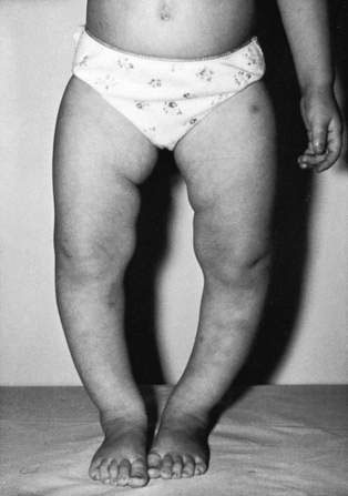
Postural talipes equinovarus
Some babies are born with one or both feet in a position of plantar flexion at the ankles, and inversion of the remainder of the foot, so that the sole of the foot faces the opposite foot. This is postural talipes equinovarus and may be distinguished from true talipes equinovarus, known as clubfoot deformity (which is rigid and not passively correctable). Postural talipes equinovarus resolves spontaneously with assistance of simple stretches and rarely a cast, if not improved at 6 weeks.
Bow legs
Bow legs (Fig. 8.1.2) are common up to 2 years of age; the parents will often be concerned that the legs are bowed and the feet turn in. The condition is not caused by bulky nappies, because the bowing is in the tibiae. It is a normal developmental process and does not require treatment apart from parental reassurance. If the bowing is in one leg only, you should investigate with plain X-rays to exclude pathological causes such as a bone dysplasia or growth abnormality.
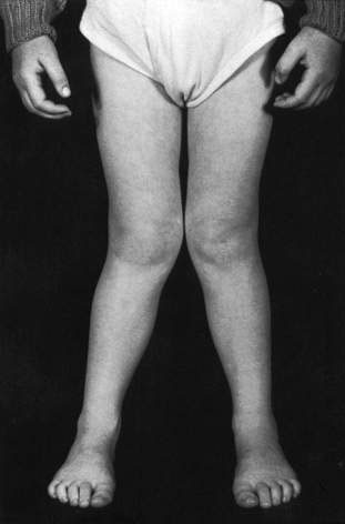
Knock knees
A large proportion of the population between the ages of 2 and 7 years have knock knees (Fig. 8.1.3). This condition has a strong tendency to correct itself by the age of 7 years and as a rule the only management necessary is parental reassurance that improvement will occur. There is a rare form of knock knees (adolescent tibia vara) that presents in overweight children around the age of 12 years and which may require treatment by staple epiphysiodesis.
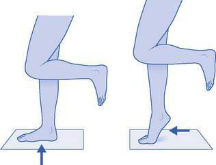
Rolling in of ankles
Parents will frequently mention this, especially after it has been noticed by a concerned grandparent or shoe-fitter. The rolling of the hind foot into valgus is due to physiological joint laxity and requires no treatment. The clinician can show the parents how the hind foot straightens when the child stands up on high tiptoes. This is the tiptoe test, which also demonstrates development of the medial longitudinal arch (Fig. 8.1.4). Orthotics for ‘rolling in’ are not required (see below).
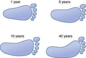
Flat feet
Flat feet in children are a frequent cause for parental concern. Usually this concern is unwarranted and the child’s foot is normal for age (Fig. 8.1.5). Often parents notice that their child’s foot appears flat. Sometimes the attendant fitter at the shoe shop may comment on the shape of the child’s foot. Children usually have low arches because they are loose-jointed and flexible. The arch flattens when they are standing. However, the arch can be better seen when the feet are hanging free or the child stands on tiptoes (the tiptoe test; see Fig. 8.1.4).
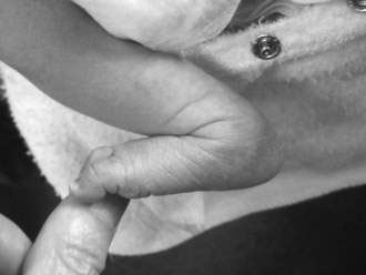
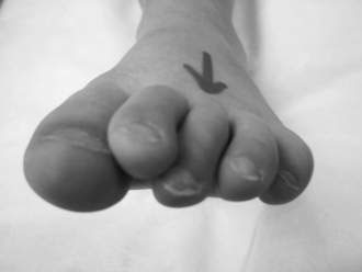
Fig. 8.1.5 Flexible flat feet are normal in infants and children. The arch develops whether the child wears shoes or goes barefoot.
When a child first learns to walk, the stance is usually wide to assist balance, and the feet roll. As the child grows and ankle muscles strengthen, the foot gradually develops its mature shape with some medial arch. Flat feet are common in preschoolers and are present in over 10% of teenagers. The final shape of the foot may also be influenced genetically, in that one or both parents may have low arches.
Between the ages of 2 and 8 years, parents are often concerned because a ‘second ankle bone’ appears on the medial aspect of the foot. They are referring to prominence of the navicular bone, which is present in most children who have flat feet. Unless the prominence of this bone is causing symptoms, it can be ignored.
Children with flat feet generally have some valgus deformity of the heel; when viewed from behind the heels do not point straight up and down, but tend to slope outwards and downwards. The heels will correct and even swing into varus during the tiptoe test. This seldom persists into adult life.
During the first 7 or 8 years of life the majority of children develop a medial longitudinal arch, but approximately 15% do not. Clearly, the results of any form of treatment for flat feet are excellent, as some 85% improve whether or not they are treated. Sometimes treatment with shoe inserts (orthotics) or other forms of arch support/shoe modification are recommended by therapists. These may satisfy concerned parents but do little, if anything, to correct the ‘flat foot’ and certainly do not make an arch where one is not present. Orthotics for flexible flat feet are not necessary for children.
Other treatments, such as splints, massage or special shoes may be offered but there is little evidence that these interventions alter the foot for the better.
Shoes
The only essential is that children’s shoes should be roomy enough. Shoes themselves are not necessary to promote normal foot growth and development; they are worn only for protection and need not be worn until activities demand this protection. Boots are no better than shoes, although parents may prefer boots for toddlers in that they are less likely to fall off or be taken off.
It is not harmful to use ‘handed down’ shoes in good condition from older children in the family, provided they are roomy enough. There is no evidence that sandals, thongs or sneakers have any harmful influence on the feet. If the child has excessive wear on the inner side of the sole of the shoes, advise parents to look for shoes that have a stiffer heel area. Some children with flexible flat feet are rather hard on their shoes and this can be dealt with by selecting shoes of stronger construction. This is usually much less expensive than elaborate and unnecessary orthotics.
Accessory navicular bone
The child with a prominent accessory navicular may have some temporary discomfort, which may be relieved by wearing arch supports for a year or two. Frequently the ossicle either unites with the main navicular bone or just becomes asymptomatic. Excision of the accessory navicular bone is required only rarely.
Curly middle toe
Sometimes the third toe curls inwards under the second toe so that the second toe tends to lie above the level of the first and third toes. Parents generally notice the abnormal posture of the second toe, but it is the third toe that is the cause of the problem. This can be safely ignored until the child is at least 2 years old. Occasionally a flexor tenotomy is required and provides excellent correction (Fig. 8.1.6).
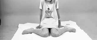
In-toe gait (pigeon toeing)
In-toeing in childhood is common. It may appear worse when the child is running or tired. It does not cause arthritis or back problems later in life. It can be due to one or more of the following:
Inset hips (persistent femoral neck anteversion) have internal rotation in excess of the range of external rotation. The condition is more common in girls and the feet seem to fly out sideways when running. The pathology lies in the top of the femur where there is a normal twist of 30° at birth, which unwinds gradually by the age of 7 years. In severe cases, when there is a major cosmetic problem unresolved by about 10 years, derotation femoral osteotomy can be performed, but this is rarely required.
Children with inset hips commonly sit between their feet with their hips in full internal rotation, the knees flexed and the legs splayed outwards (the ‘W’ position) (Fig. 8.1.7). This is the only way they can sit comfortably as they cannot externally rotate their hips sufficiently to sit in a cross-legged fashion. It is almost unknown for an adult to present with a complaint of in-toeing, which tells us that the natural history is spontaneous resolution.
Internal tibial torsion (a twist in the shin bone) is usually due to intrauterine pressure and can persist up to the age of 3 years and then spontaneously corrects.
Metatarsus adductus (Fig. 8.1.8) is a condition in which the feet are banana-shaped, with the convexity of the banana outwards and the toes directed towards each other. This may be due to intrauterine pressure; however, if it persists it is called metatarsus adductus. It is passively correctable and slowly rights itself, especially after walking commences. Very rarely, manipulation and plaster immobilization is necessary.
Congenital abnormalities
Developmental dysplasia of the hip
This condition was previously called congenital dislocation of the hip (CDH); however, developmental dysplasia of the hip (DDH) is now the preferred term as it tells us that some of these hip problems develop after birth. DDH is the most common musculoskeletal abnormality in neonates. The incidence of this condition in Australia and North America is 7 per 1000 live births. In some regions of Europe it is more common.

Full access? Get Clinical Tree

