Oral Pathology
Andrew L. Sonis and Brian Grove
While dental caries and its consequences represent the most common oral pathology encountered in children, both localized and systemic conditions of noncariogenic origin may manifest themselves in the mouth and surrounding tissues. This chapter will review some of the common and uncommon pathologies observed in or around the oral cavity of children.
SOFT TISSUE PATHOLOGY
 ANGIONEUROTIC EDEMA
Angioneurotic edema is an allergic reaction often involving the oral soft tissues (see Chapter 189). Antigens precipitating this reaction include food and latex allergies, vaccines, insect bites, and medications. Typically, facial edema follows exposure to the antigen and may include swelling of intraoral soft tissues, potentially resulting in airway embarrassment. Initial management is provided with antihistamines. More aggressive therapy may require the administration of corticosteroids.1–10
 ANKYLOGLOSSIA
Ankyloglossia (tongue-tie; Fig. 378-1) is a congenital anomaly of the tongue characterized by a short and sometimes anteriorly inserted frenulum. The clinical significance of ankyloglossia is a matter of controversy, particularly as it relates to breastfeeding difficulties such as sore nipples, poor infant weight gain, neonatal dehydration, and shortened breastfeeding duration. Infants with ankyloglossia and associated problems with latching may benefit from frenectomy. Rarely in the older child will it interfere with speech or contribute periodontal problems. Recent studies suggest some advantage of laser frenectomy over traditional surgical frenectomy relative to pain and recovery times.11–13
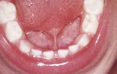
FIGURE 378-1. Ankyloglossia (tongue tied). A short lingual frenum may interfere with feeding or speech and may necessitate surgical intervention.
 BACTERIAL INFECTIONS
Dental caries is the most common bacterial infection in the mouth that may progress to abscess and cellulitis if left untreated. (The caries process is described in Chapter 374). Non-tooth-related intraoral bacterial infections are relatively uncommon. Extraoral bacterial infections tend to be far more common (see Chapter 367). Several other systemic conditions that have bacterial etiologies can present with oral lesions. These include streptococcal infections (see Chapter 285), diphtheria, tuberculosis, cat-scratch disease, actinomycosis, gonorrhea, and syphilis.
 BITE INJURIES
Accidental self-inflicted bite injuries are quite common in children, especially after having received intraoral local anesthesia. Not infrequently, children who have received a mandibular nerve block for dental treatment will chew their lip, buccal mucosa, or inner lip unknowingly, resulting in a significant area of epithelial necrosis accompanied by edema. Sites most frequently involved are the lateral border and tip of the tongue, buccal mucosa, and lower lip. The injury usually presents as a macerated, irregular lesion with painless white necrotic areas of epithelium surrounding the ulcerated surface. Treatment, if necessary, is supportive and palliative since these areas of trauma rarely, if ever, become secondarily infected.17
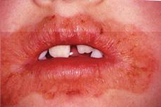
FIGURE 378-2. Cheilitis. The habit of chronic lip-licking can result in significant inflammation of the perioral tissues. Severe cases typically respond well to topical steroids.
 CHEILITIS
Cheilitis (Fig. 378-2) is a common inflammatory disorder in children. It typically arises from the chronic habit of licking the lips and perioral skin. Less frequently, cheilitis or angular cheilosis (perlèche) may occur from mechanical irritation, malocclusion, atopic dermatitis, fungal infection (typically candidiasis), bacterial infection, or nutritional deficiencies. The clinical features of cheilitis are erythema, mild edema, and thin scaling formation of the lips and perioral areas. When due to chronic lip-licking, treatment should be directed at breaking the habit. Empirically, this may be accomplished with lip balms with undesirable tastes. In severe cases, topical steroids, with or without fungicidal and bactericidal activity, may be beneficial. Obviously, in cases where a nutritional deficiency is thought to be the underlying etiology, nutritional supplements are indicated.18-20
 CONGENITAL EPULIS
Congenital epulis (Fig. 378-3) or gingival granular cell tumor of the newborn is a relatively rare lesion typically arising from the maxillary anterior alveolus. Far more common in females (8:1), it presents as a smooth, firm, pedunculated soft-tissue lesion attached to the alveolus by a broad base. It can range in size from several millimeters to several centimeters, with larger lesions frequently being detected in utero.
 ERUPTION HEMATOMA
An eruption hematoma (Fig. 378-4) is a soft tissue variant of the dentigerous cyst that develops around the crown of an erupting primary or permanent tooth. It is quite common and can occur in association with any erupting tooth. The lesion typically resolves on its own with the eruption of the underlying tooth. Rarely is it necessary to excise the roof of the cyst to allow for eruption of the involved root.
 FIBROMA
The irritation fibroma is considered the most common childhood reactive lesion of the oral cavity. In the vast majority of cases, the lesion arises as a reaction to a local irritant. While the lesion may involve any oral anatomic site, the buccal mucosa and interdental gingiva are the most common sites in children. The lesion usually presents as an asymptomatic pedunculated or sessile growth secondary to irritation. Treatment is surgical excision with the elimination of any associated source of local irritation.22,23 Failure to remove the source of irritation will likely result in recurrence.
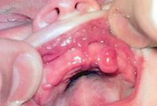
FIGURE 378-3. Congenital epulis. These benign neoplasms typically arise from the anterior maxilla and can range in size from several millimeters to centimeters. Excision of smaller lesions not interfering with function may be deferred several months to minimize anesthetic risk.
 CANDIDIASIS
Candidiasis or candidosis is an infection with the yeastlike fungal organism Candida, the most common being Candida albicans. It often infects children and adults who are immunocompromised or who are on a prolonged course of antibiotic therapy. It is the most common oral fungal infection in humans and can be manifest in a variety of clinical presentations, which may make appropriate diagnosis difficult.24
Erythematous, or chronic atrophic candidiasis, may also be caused by immunosuppression, xerostomia, or chronic exposure to broad-spectrum antibiotic therapy. Patients often complain of a burning sensation on their tongue associated with a loss of filiform papilla and a denuded appearance of the tongue’s dorsal surface. Unlike the acute form characterized by sloughing white lesions, the erythematous form results in patchy lesions. Many forms of chronic candidiasis are asymptomatic, including central papillary atrophy and median rhomboid glossitis. In addition, angular cheilitis is a particular form of erythematous candidiasis that affects the commissures and is characterized by fissuring, erythema, and scalelike lesions. The treatment regimen is similar to that of acute candidiasis.24–27
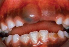
FIGURE 378-4. Eruption hematoma. Often associated with an erupting primary tooth, these benign lesions are the result of hemorrhage into the cystic cavity surrounding the crown of the involved tooth.
Pseudomembranous candidiasis (acute) is the most common form of oral candidiasis, which is usually seen on the buccal mucosa, dorsum of the tongue, and soft-palate (eFig. 378.1  ). It is commonly known as thrush and appears as white cottage cheese–like lesions that can be removed by scraping. It is the most common manifestation of HIV infection in children. Topical antifungal medications are often the drug of choice for localized candidiasis. While compliance for the “swish and swallow” routine with these medications is readily accomplished by older patients, in the infected infant, appropriate delivery of the topical medication is often problematic. A foam-headed device (Toothette) is a useful aid in applying the medication to the infected oral mucosa. Pharmacotherapy includes Nystatin oral suspension, Clotrimazole troche, or systemic antifungal mediations such as ketoconazole or fluconazole.24–27
 GEOGRAPHIC TONGUE (ERYTHEMA MIGRANS)
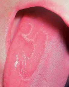
Geographic tongue (Fig. 378-5) is a harmless common disorder of unknown etiology that primarily affects the dorsal surface of the tongue, although it may occasionally involve other mucosal surfaces, including the buccal and sublingual mucosa. The involved area typically changes or “migrates” over several weeks or months, thus giving rise to the disorder’s name. Onset tends to be fairly rapid, with lesions typically persisting for several weeks, although occasionally they may last for several years. Patients are generally asymptomatic but may rarely complain of a “burning” sensation. No treatment is necessary; however, patients experiencing burning may benefit from palliative treatment with topical anesthetics.28
Stay updated, free articles. Join our Telegram channel

Full access? Get Clinical Tree


