Office Evaluation of the Eyes
Gregg T. Lueder
OCULAR HISTORY
The appropriate ocular history will vary depending on the reason the child has come for evaluation. If a child presents for a routine, well-child examination, the ocular history will be included in the review of systems. The caregiver should be asked about any specific eye complaints. In infants and younger children, the examiner’s questions should include whether the child appears to see normally, if there are any abnormal eye movements, or if there are any visible abnormalities of the eyes or periocular structures. Older children may be questioned directly regarding subjective symptoms, including blurred vision (difficulty seeing the blackboard at school or objects that friends or family members can see easily), double vision, or other concerns regarding vision or the appearance of the eyes.
If a child presents for evaluation of an ocular problem, the appropriate history will depend on the specific complaint. As with any medical concern, questions should include the onset and duration of symptoms, aggravating and alleviating factors, history of similar problems, and previous treatment.
General medical and family histories are also important in evaluating the eyes. Children who are born prematurely or who had perinatal difficulties have an increased risk of developing vision problems. Many systemic disorders, such as trisomy 21 and Marfan syndrome, are associated with specific ocular problems. Children with juvenile idiopathic arthritis are at increased risk for asymptomatic intraocular inflammation (uveitis) and therefore require longitudinal screening.1 Children with neurofibromatosis type 1 require annual examinations at least during the first 7 years of life to identify signs of optic pathway glioma. A family history of an ocular condition or systemic disease that affects the eye may also alert the examiner to look for specific problems. If family members have been affected by serious eye disorders that may be inherited, such as infantile cataracts, pediatric glaucoma, or retinoblastoma, screening by an ophthalmologist may be indicated, even if the office evaluation appears normal. More common familial problems include strabismus, myopia, and astigmatism.
When obtaining an ocular history, it is important to recognize that children may be unaware that they have a vision problem, even with significant visual impairment. Young children in particular typically adapt very well to decreased vision and often do not voice complaints, even when vision is below the level of legal blindness (worse than 20/200 in the better eye or severe visual field constriction). The visual demands of infants and toddlers are relatively minor—they only need to see well enough to find their food and toys and identify family members, and they do not need to read fine print. Therefore, they may appear to function quite well, often until grade 1 or 2, when visual demands begin to increase. In addition, children whose vision loss is confined to one eye also frequently do not voice complaints, even if specifically questioned. They function well with the vision in the normal eye and usually ignore the eye with the problem. This ability to unconsciously “ignore” one eye is the basis for the development of amblyopia. Even older children may be completely unaware of chronic unilateral vision impairment until the eyes are tested independently. For these reasons, the absence of vision complaints in children does not adequately rule out vision problems. The American Academy of Pediatrics recommends vision screening at every well-child visit.2 In addition to assessing vision, the pediatrician should be skilled at basic complete eye examination, to be used as time permits, as circumstances indicate, and as specific complaints or concerns may require.
OCULAR EXAMINATION
Before children are old enough to read an eye chart, vision is assessed by watching the child fixate on objects, observing whether the eyes track smoothly and assessing whether the eyes move together equally. Although children with developmental delay or neurological abnormalities may be less attentive, more distractible, or difficult to engage, one should not assume that poor responsiveness to a visual target is due solely to the neurological condition. Consultation with an ophthalmologist may be needed to specifically assess the vision. The degree of interest the child has in these activities will be variable. Some children enjoy the examination, viewing it like a game, while others are apprehensive and have difficulty cooperating regardless of how nonthreatening the assessment may be. In addition, children’s level of interest may change depending on whether they are awake and alert or tired and irritable, and whether they are feeling well in general. If the child is uncooperative due to identifiable reasons that are likely to resolve and there are no vision or ocular concerns, it may be reasonable to defer the vision screening to a subsequent visit, but the pediatrician must be sure that this follow-up occurs.
It is useful to have brightly colored, interesting objects to hold in the examiner’s hand to monitor fixation and tracking. However, children may quickly become bored with a single toy, so it is useful to have a few toys that can be interchanged. In addition, it is helpful if the examiner can be flexible regarding the order in which the examination is performed. Looking at toys while sitting in their parent’s lap bothers few children, but they may begin crying or fighting if the examiner attempts to touch an area near their eyes. Therefore, it is best to perform the least invasive portions of the examination first, leaving the potentially more bothersome portions to the end. Although the specifics of the examination will be presented below, beginning with the front of the eyes and moving backward, the order may be adjusted to the child’s cooperation.
ASSESSMENT OF VISION
Although assessing vision may sometimes be difficult in young children, it is critically important to identify problems as early as possible. Vision matures rapidly in the first few years of life, and interruption of this process may produce irreversible loss of vision due to amblyopia. Severe problems, such as unilateral infantile cataracts, need to be corrected within the first few months of life if vision is to be restored. The prognosis for treatment of amblyopia due to strabismus or unequal refractive error is related to the age at which the problem is identified and treatment begun. The younger the child, the better the prognosis. However, it is also more difficult to assess vision in younger children.
The method of vision assessment varies with the patient’s age. Normal infants can fixate at birth, in particular on their mother’s face. This response is well recognized by parents feeding their child. Asking the parents of an infant, especially when they have previously raised a normal child, whether their child sees is a remarkably accurate assessment of the child’s vision. In the first 1 to 2 months of life, infants should at least respond to lights by blinking when a bright light is shone into the eyes. When the lights are turned off, the eyes often open, sometimes with the upper lids retracting such that the superior sclera (white of the eyes) becomes visible, a primitive response known as the eye-popping reflex. Many infants will track fairly well shortly after birth, but it is not abnormal to have minimal tracking at this early age. A 3-month-old infant should be able to fixate on a toy held in the examiner’s hand and should track the object back and forth as the examiner moves it. This is initially done with both eyes open. However, because an infant may track well if the vision is good in only one eye, it is important that the vision in the two eyes be assessed independently. This is done by covering each eye separately (usually with the examiner’s or caretaker’s hand) and monitoring whether the infant tracks equally well with either eye. If the child consistently tracks well with one eye covered but becomes upset or refuses to track when the opposite eye is covered, this strongly suggests that there is a vision problem in the first eye that was covered. An important caveat, however, is that some children get upset when either eye is covered, even if both eyes see well. Therefore, if the child becomes agitated equally with either eye covered, one may not be able to accurately judge the vision based on this behavior. In addition to distracting the child with interesting toys as vision stimuli, it is also important to try avoiding direct contact with the child’s face when occluding one eye. This can be achieved by placing the examiner’s hand slightly in front of, but not directly in contact with, one side of the face. Also, the examination should be conducted fairly quickly. If there is a difference in the visual responsiveness of one eye versus the other, it will often be readily apparent.
As children become older, usually by age 3 years, testing of vision with eye charts is possible. The child is placed a standard distance from a chart, the eyes are covered one at a time, and the child is asked to describe what they see. A variety of charts are available for use in the pediatric setting. In younger children, charts with readily identifiable symbols or pictures are usually most effective. The tumbling E test may also be used, but children may have more difficulty understanding this test, and before the age of 4 to 6 years, handedness may not be sufficiently developed to allow full compliance. Some children will be hesitant to vocalize what they see. Matching games or cards can be used, such as the HOTV test, that allow the child to point to a handheld card to match what they see, rather than speak out loud. By age 5 years, most children can read an adult eye chart with letters. Specific guidelines that include the details of how to perform these tests and the criteria for referral have been published by the American Academy of Pediatrics.2
Assessment of color vision may also be included in the ocular examination, but this is not required. Most children with color vision deficits function entirely normally and may not even realize they have a deficit until adulthood. Deficit in the red-green color system is an X-linked recessive disorder that is present in approximately 8% of males and 1% of females. It is important for families and teachers to be aware of this disorder so the etiology of an affected child’s inability to distinguish colors is properly identified. Tests such as the Ishihara test plates are used to screen for red-green color deficiency by having the individual identify colored numbers within different colored backgrounds. This test can normally be performed by age 5 to 6 years. If there is parental or teacher concern about color discrimination beyond this age, referral to an eye specialist may be indicated.
Binocular vision and depth perception may be affected by amblyopia, strabismus, and monocular visual impairment. However, children rarely experience significant functional problems due to these deficits. Formal testing usually requires consultation with an ophthalmologist. In particular, dyslexia and other reading problems are rarely due to ocular problems such as binocular vision or tracking difficulty. An ophthalmic examination is indicated in children with reading difficulties to rule out significant refractive errors or strabismus that could interfere with reading. In the absence of such problems, appropriate intervention includes remedial educational programs directed to reading strategies, rather than vision therapy, for which there is no scientific evidence of efficacy.3
EYELIDS, PERIOCULAR STRUCTURES, AND LACRIMAL SYSTEM
The eyelids in children should be symmetric, and the margin of both upper eyelids should rest above the pupil. If one or both eyelids droop (ptosis), children may adopt a chin-up head posture to view below the drooping eyelid(s), or they may contract the forehead frontalis muscles to assist in elevating the lid by raising the brow. Therefore, when evaluating eyelid height and function in a patient in whom ptosis is suspected, the examiner must be sure that the child’s head is in normal position and the frontalis muscles are not contracting. If the eyelid in an infant is covering most of the eye, particularly the pupil, prompt ophthalmologic evaluation is indicated because of the risk of permanent vision loss due to amblyopia.
The eyelid margins should be continuous and should rest against the globe, and the eyelashes should turn outward and not rub against the eye. The skin of the eyelid is very loose, and any condition that causes edema often manifests relatively early by swelling of the lids. Eyelid edema is usually most prominent upon awakening, due to the effects of gravity during sleep. This can be aggravated by vascular abnormalities such as port-wine stains. Likewise, following trauma or surgery, ecchymosis can accumulate quite extensively in the eyelids and may even make it difficult to open the eyelids.
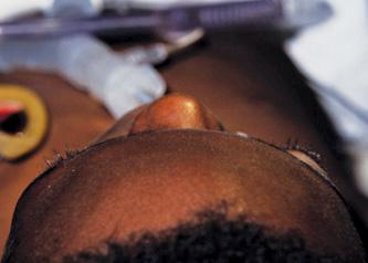
Inflammation or masses in the eyelid or periocular structures should be noted. If an orbital mass or hemorrhage is present, patients may present with proptosis. In this condition, the entire eyeball is pushed forward and appears to bulge from the orbit. Widening of the space between the eyelids due to this bulging produces an asymmetric appearance, which is sometimes mistaken as ptosis of the normal eye. The proptotic eye appears more prominent and is often best assessed by looking down from above the patient (Fig. 580-1). The eye may be irritated due to corneal exposure and incomplete eyelid closure. Proptosis may be caused by several disorders and requires prompt evaluation by an ophthalmologist.
CONJUNCTIVA AND ANTERIOR SEGMENT
The conjunctiva and anterior segment are best examined with a penlight or the light from a direct ophthalmoscope. If a more detailed view is required, the examiner can look through the direct ophthalmoscope, adjusting the dial on the instrument to provide a focused, magnified view. If an infant or child is not anxious and the light is not too bright, this examination can be performed while the patient is sitting in the caretaker’s lap. If the eyelids are squeezed shut, they may need to be manually opened in order to perform the assessment. This can usually be done with the examiner’s fingers, but use of cotton-tipped swabs or an eyelid specula is sometimes necessary. The conjunctiva in newborns may have a yellowish tinge due to elevated bilirubin levels. Irritation of the conjunctiva may produce edema (chemosis). Chemosis is also seen after craniofacial surgery and in association with severe systemic edema. A red eye is due to infection of the conjunctival blood vessels. Smoke and chorine in swimming pools are common sources of irritation. Conjunctival swelling and infection associated with discharge are common manifestations of conjunctivitis. Conjunctival inflammation may also occur with marked increase in intraocular pressure (glaucoma) or iritis.
Stay updated, free articles. Join our Telegram channel

Full access? Get Clinical Tree


