Nongynecologic Conditions Encountered by the Gynecologic Surgeon


Nongynecologic Conditions Encountered by the Gynecologic Surgeon


Kellie L. Mathis

William A. Cliby


INTRODUCTION

In the majority of patients proceeding to the operating room for surgery in the pelvis, an accurate diagnosis has been made preoperatively. Advances in imaging should reduce the number of unanticipated findings intraoperatively for the pelvic surgeon. But, occasionally, the gynecologic surgeon will encounter an unexpected finding in the abdomen or pelvis intraoperatively arising from outside of the reproductive tract. This is commonly a problem in obese patients and elderly patients with atypical or ill-defined pelvic pathology and requires methodical consideration of the differential diagnosis preoperatively to appropriately triage and plan for these unanticipated findings. We discuss the most common nongynecologic findings in this chapter.


PREOPERATIVE EVALUATION

A thorough history and physical exam is absolutely critical. This should include presenting symptoms and past medical history as well as family history. The examination should include a thorough pelvic exam as well as a digital rectal examination. If the patient has any gastrointestinal complaints, a fecal occult blood test should be performed as well. Perianal abnormalities such as fistulae, atypical fissures, and large skin tags should raise the suspicion of Crohn disease.

If the patient complains of abdominal pain, low-grade fevers, changes in bowel habit, or blood in the stool, then a complete gastrointestinal workup should precede surgery. This may require endoscopy as well as computed tomography (CT) imaging. CT enterography should be ordered when there is a concern for small-bowel disorders. Patients suspected of having gynecologic infections and possible tuboovarian abscesses should be screened for sexually transmitted diseases.

If the patient has urinary symptoms, a urinalysis and culture should be done. If there is evidence of hematuria and/or bacteria, the patient should be treated for a urinary tract infection and reevaluation done to confirm clearance. If hematuria persists, further urinary workup should be considered to evaluate the upper urinary tracts as well as rule out intrinsic bladder pathology. This may include CT imaging, cystoscopy, or renal ultrasound.


PREOPERATIVE PREPARATION

The most important preoperative consideration is to make an accurate diagnosis or when not possible to define the differential diagnosis so the appropriate surgical expertise can be involved in the case. If there is any uncertainty about the diagnosis, an extensive discussion with the patient and informed consent about possible findings and interventions should occur. If there is any suspicion that intestinal surgery may be necessary, then the patient should be informed that a protective ostomy may also be necessary, and the abdomen should be marked preoperatively by a wound ostomy nurse. Emerging evidence suggests that postoperative complications are less common when a wound ostomy nurse sees, marks, and educates the patient preoperatively. Other subspecialists should meet and counsel the patient as appropriate. Other principles of safe surgery should be followed, including ensuring adequate nutritional and hydration status. Mechanical bowel preparation is no longer recommended or necessary for safe intestinal surgery. If the surgeon anticipates possible leftsided colon resection, and most commonly for pelvic pathology where pelvic colon involvement is always a possibility, we recommend that the patient be given two tap water enemas upon admission on the morning of surgery. The rectum should also be irrigated with dilute Betadine just before prepping the abdomen and perineum.


INTRAOPERATIVE FINDINGS AND MANAGEMENT

The patient should be placed in the modified lithotomy position, and a sterile Foley catheter should be placed. Appropriate antibiotics timed correctly should be given, and in most cases, venous thromboembolism prophylaxis would be recommended according to current standards of care. Ureteral stents should be considered, especially in the setting of multiply recurrent surgeries and large pelvic masses. Consider a minimally invasive approach (laparoscopic/robotic) to perform the operation, or in cases of uncertain etiology, a diagnostic laparoscopy can be performed to aid in the diagnosis or incision planning or to determine if triage to a higher-level care center is necessary. If an open approach is followed, careful placement of the abdominal incision is essential to ensure that the upper abdomen can be reached if necessary; a midline incision is preferred, particularly when the diagnosis is uncertain. Adhesiolysis should be performed first if necessary followed by a full abdominal exploration. Self-retaining retractors should be placed to minimize the risk of neurologic injury, particularly femoral nerve injuries, and avoid undue tension on the abdominal muscles. If the small intestine is uninvolved, then it should be packed away into upper abdomen. Generally, the patient is put in the Trendelenburg position, and the pelvis should then be assessed.

We now consider some of the nongynecologic pathology that may be encountered in the abdomen and pelvis.


GASTROINTESTINAL DISORDERS


Appendicitis

If appendicitis is in the differential diagnosis preoperatively, laparoscopy should be performed. A 12-mm Hasson trocar should be placed at the umbilicus with two additional 5-mm trocars in the suprapubic midline and left lower quadrant. The patient should be positioned with the right side up. If the appendix is grossly inflamed and dilated in the absence of other pathology, the appendix should be removed. The appendix should be gently grasped with a Babcock clamp. The mesoappendix can be transected using hemostatic clips or vessel-sealing device or with an endoscopic linear stapler. A stapler should be used to transect the appendix at the base of the cecum. The specimen can then be pulled through the umbilicus in a bag or glove to avoid wound contamination. If there is gross purulence due to perforation, the area should be copiously irrigated with saline prior to closure. If a phlegmon is encountered in the right lower quadrant and the cecum is involved, it is reasonable to abort the procedure and treat the appendicitis with intravenous antibiotics and percutaneous drainage if an abscess develops. Surgery in this setting is associated with higher morbidity. If an organized abscess is seen, placement of a drain at the time of surgery is suggested; in the case of a phlegmon, no drain is recommended.


Diverticular Disease

Diverticulosis is common among Americans, with prevalence rates up to 45%. Diverticulitis is inflammation of the diverticulum, usually occurring in the sigmoid colon, and it is generally managed nonoperatively. Uncomplicated diverticulitis accounts for 75% of all cases and is initially treated with bowel rest, antibiotics, and possible hospitalization (
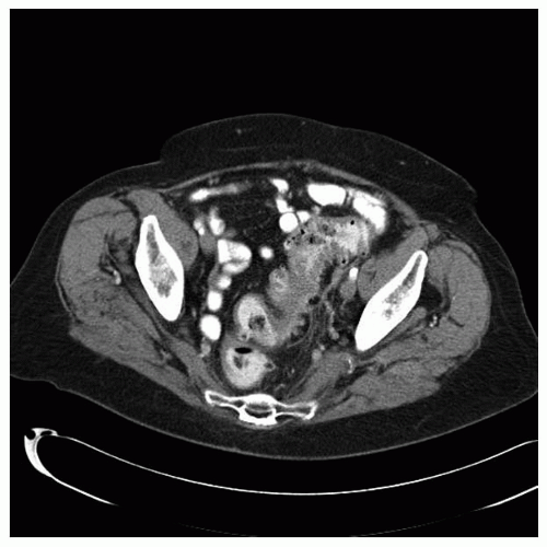
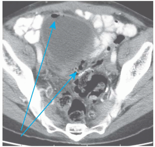
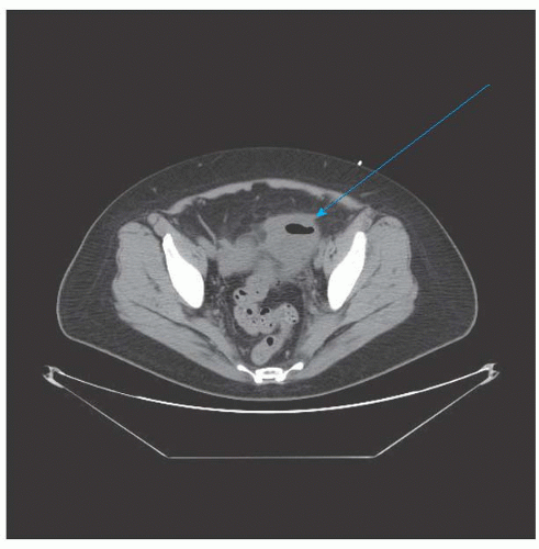
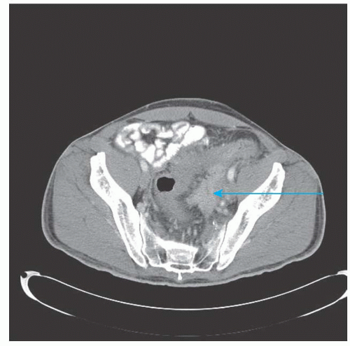
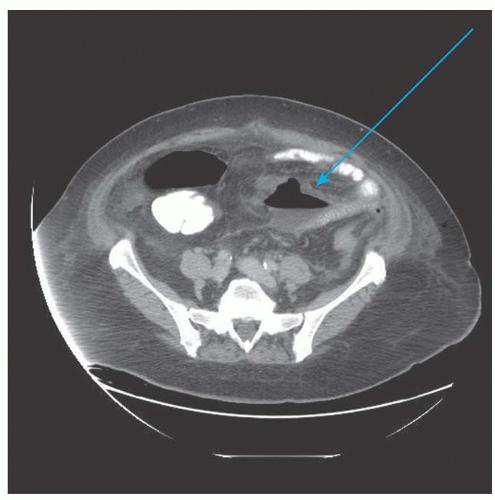
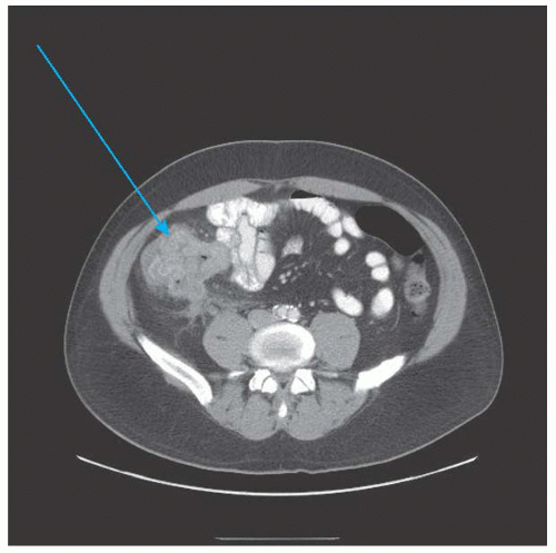
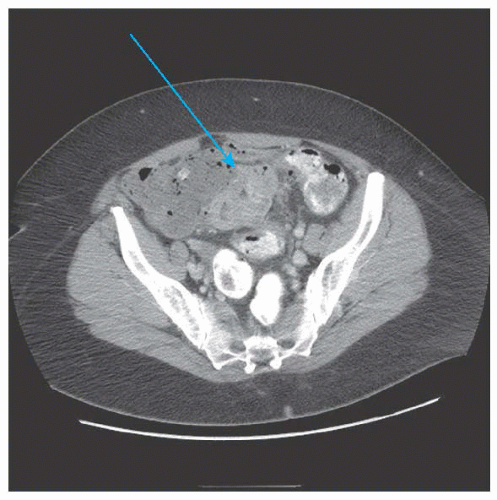
Fig. 48.1). Only when the attacks become recurrent and frequent enough to affect lifestyle is elective sigmoid resection entertained. Complicated diverticulitis (presence of abscess, perforation, fistula, or stricture) generally requires surgery (Figs. 48.2,48.3 and 
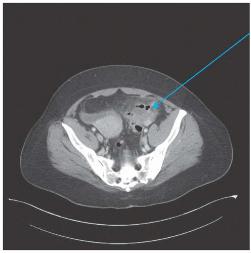
48.4). These cases often present to, or are referred to, a gynecologist because pain localizes to the pelvis and imaging demonstrates a pelvic mass, so a high index of suspicion is required when evaluating patients with preoperative gastrointestinal symptoms or systemic signs of infectious etiologies. When a contained abscess is seen on CT imaging, percutaneous drainage is attempted (Fig. 48.5). Surgery is ideally performed a few weeks after drain removal in an effort to avoid an end or diverting stoma. Patients with an acute perforation will present with an acute abdomen and require emergency surgery (Fig. 48.6).


FIGURE 48.1 Uncomplicated sigmoid diverticulitis.


FIGURE 48.2 Sigmoid diverticular disease with colovesical fistula (note pericolic gas and gas in bladder [arrows]).


FIGURE 48.3 Complicated diverticulitis versus tuboovarian abscess (arrow).


FIGURE 48.4 Perforated diverticulitis with abscess involving left adnexa (arrow).

If inactive diverticulosis is found intraoperatively, it should be ignored. Diverticulosis is common, and the majority of patients will never require resection. If the patient has acute diverticulitis or chronic smoldering diverticulitis, it is reasonable to consider a sigmoid resection. The technique of sigmoid resection is outlined in the Steps in the Procedure box.


FIGURE 48.5 Acute sigmoid diverticulitis with a contained abscess (arrow).


FIGURE 48.6 Acute diverticulitis with noncontained perforation (arrow).


STEPS IN THE PROCEDURE


Sigmoid Resection for Diverticulitis


  • Retract the sigmoid colon medially and separate the colon from the white line of Toldt and Gerota fascia. Identify and protect the left ureter and ovarian vessels.


  • Mobilization of the splenic flexure may be necessary if there is not a natural redundancy in the colon.


  • Divide the mesentery to the sigmoid colon, staying close to the bowel wall. This is done with right angle clamps and ligatures in the open technique and with a vessel-sealing device in the laparoscopic technique.


  • Identify and transect the upper rectum with a TA stapler.


  • Identify and transect the descending colon. Secure the anvil of the circular stapler into the proximal lumen with a purse-string suture.


  • Perform a tension-free end-to-end stapled anastomosis using a circular stapler.


In order to avoid recurrence of diverticulitis, it is critical that the surgeon resects the entire sigmoid colon and perform the anastomosis between the soft and nonedematous descending colon and the upper rectum. It is not necessary to resect all of the diverticula from the proximal colon. Mobilization of the splenic flexure may be required to perform an anastomosis without tension. If the setting of significant inflammation around the new connection, it is prudent to consider a temporary proximal diversion. If the tissue quality is poor (due to inflammation, edema, size mismatch, etc.), a Hartmann procedure is the safest approach.

Diverticulitis with an abscess involving the adnexa can sometimes be mistaken for a tuboovarian abscess, and this should always be in the differential diagnosis of a pelvic mass in the older female patient. In such cases, moderate elevation of CA-125 is common secondary to inflammation, which can further complicate the diagnostic challenge.


Colorectal Cancer

Cancers of the colon are common with over 100,000 new diagnoses in the United States every year. Ninety percent of colon cancers occur after the age of 50, but they can occur in every age group. Surgical resection is the primary treatment colon and rectal cancer. If a cancer of the colon is unexpectedly encountered intraoperatively, a decision should be made as to whether to proceed with bowel resection immediately or close the abdomen and perform the definitive surgery at a later date. If an operating surgeon experienced in oncologic colon resections is immediately available, then resection and primary anastomosis can be done. As stated before, mechanical bowel preparation is not necessary, but enemas are generally given preoperatively to empty the colon of the bulk of the stool for a left-sided resection. The most frequent tumors unexpectedly encountered by the gynecologic surgeon are cecal (Figs. 48.7 and 48.8) or rectosigmoid in location. Due to locations, these can often be mistaken for an adnexal mass. Techniques for each are described in the “Steps in the Procedure” boxes. The liver and peritoneal surfaces should be closely inspected for metastatic lesions prior to closure of the abdomen.


FIGURE 48.7 Advanced cecal cancer (arrow).


FIGURE 48.8 Large cecal cancer with local invasion into the sigmoid colon (arrow).


STEPS IN THE PROCEDURE


Right Colectomy Technique for Cecal Cancer


  • Explore for resectability (omentum, peritoneum, liver, duodenum, superior mesenteric vessels).


  • Retract the colon medially and separate the colon from the white line of Toldt and Gerota fascia. Identify and protect the ureter, gonadal vessels, and duodenum.


  • Dissect the gastrocolic ligament to complete the mobilization of the hepatic flexure.


  • Identify the appropriate vessels. Divide the ileocolic, right colic, and right branch of the middle colic vessels near their origins.


  • Complete the mesenteric dissection. There is an expectation that at least 12 lymph nodes (LNs) are harvested because of a known survival advantage associated with increasing number of LNs.

    Only gold members can continue reading. Log In or Register to continue

    Stay updated, free articles. Join our Telegram channel

    Jun 4, 2016 | Posted by in GYNECOLOGY | Comments Off on Nongynecologic Conditions Encountered by the Gynecologic Surgeon

    Full access? Get Clinical Tree

    Get Clinical Tree app for offline access