Fig. 7.1
Nevus of Ota affecting the temple, maxillary area, and sclera of the skin
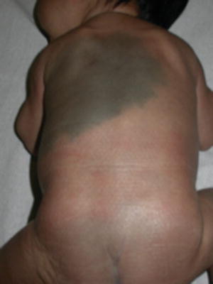
Fig. 7.2
Nevus of Ito
Epidemiology
Nevus of Ota and nevus of Ito are sporadic
They are more common in Asian and African-American children
Nevus of Ota is five times more common in females.
Nevus of Ota and nevus of Ito are sporadic lesions more common in Asians and African Americans and are up to five times more common in females [2, 3]. The incidence of nevus of Ota in darker skin has been estimated to be around 0.016 % [4]. Nevus of Ito is less frequent and the exact incidence is unknown.
Clinical Features
Nevus of Ota and nevus of Ito are speckled, irregular blue-gray macules in a characteristic distribution
Nevus of Ota affects the ophthalmic and maxillary branches of the trigeminal nerve including the sclera of the eye
Nevus of Ito affects the posterior supraclavicular, scapular, or deltoid regions
They may be present at birth or appear later
They may be associated with capillary malformations (port-wine stains) in phakomatosis pigmentovascularis type II
Nevus of Ota and nevus of Ito present as bluish to gray irregularly shaped and sometimes speckled macules. They are usually present at birth, although in as much as 40 % of cases the onset is at puberty [1, 3]. Nevus of Ota usually affects the periorbital region, temple, forehead, malar region, and/or nose. The ipsilateral sclera may be pigmented in two-thirds of cases and may also affect the ipsilateral iris, conjunctiva, retina, cornea, choroid, extraocular muscles, and retrobulbar fat. Oral mucous membranes and palate may be involved. It is usually unilateral though at times bilateral cases may occur. The nevus of Ito involves the area supplied by posterior supraclavicular and lateral cutaneous brachial nerves which are the side of the neck, the supraclavicular and scapular areas, and shoulder region. Nevus of Ota nevus of and Ito are usually unilateral, but they may be bilateral and coexistence of nevus of Ito and Ota in the same patient has been described [5].
Nevus of Ota and Ito may also coexist with capillary malformations in the so-called phakomatosis pigmentovascularis or cesioflammea. Coexistence with nevus spilus has also been reported [6].
Pathogenesis
It is believed that nevus of Ota and nevus of Ito are the result of a failure of migration of the melanocytes from the neural crest to the epidermis. They are histologically characterized by the presence of melanin-producing dendritic melanocytes within the upper and mid-dermis with their axis parallel to the skin surface. The overlying epidermis is normal. Recently mutations in GNAQ have been detected in some and other congenital dermal melanocytosis [7].
Treatment
Q-switched laser treatment of nevus of Ota and Ito is possible.
Many treatments are necessary and repigmentation may occur.
Treatment of nevus of Ota and Ito may be considered for cosmetic reasons, particularly if the patient is bothered by the appearance of the lesion. They may be treated with Q-switched ruby, alexandrite, or Nd:YAG lasers [8–10]. Multiple sessions in the range of 7–10 or more are usually needed. Repigmentation after years is the main caveat. The earlier the treatment is given the more rapid clearing but also the higher recurrence rate. Dermabrasion and cryotherapy have also been used although there is a risk of scarring and therefore are not generally recommended [11, 12]. Dyspigmentation in the form of hyperpigmentation and/or hypopigmentation can occur with laser therapy in patients of color, especially children with Fitzpatrick types IV or greater.
Stay updated, free articles. Join our Telegram channel

Full access? Get Clinical Tree


