Musculoskeletal Emergencies
Martin G. Hellman
As a primary care provider, a pediatrician should have a fundamental knowledge of musculoskeletal injuries and fractures in children. Of all childhood injuries, 10% to 15% are musculoskeletal. An understanding of the unique nature of pediatric fractures and their acute care is important to every pediatrician. Recognizing fractures secondary to possible child abuse is critical.
PHYSIOLOGIC HIGHLIGHTS OF CHILDREN’s BONES
Several key points concerning the anatomy of children’s bones must be emphasized.
The growth plate (physis) is susceptible to injury because of its relative weakness. The physis, located at the end of long bones, is under compressive forces, and is radiolucent. Ligaments at joints are stronger than the physis until it closes at puberty; therefore, true sprains are rare or nonexistent. An apophysis is a bony prominence to which muscles or tendons are attached. It is joined to the bone by a radiolucent physis that is under traction forces.
Blood is supplied to the epiphysis by penetrating vessels from the joint capsule, whereas the metaphysis and diaphysis of the bone receive blood from nutrient and periosteal vessels. The physeal blood supply is from epiphyseal, metaphyseal, and perichondrial vessels. Fractures to the growth area can cause vascular compromise and subsequent growth problems. The periosteum of children’s bones is thicker, stronger, and more vascular, but less attached than that of adult bones. Because of these properties, callus forms more quickly in children, and limits the amount of displacement associated with fractures.
Children’s bones are different biomechanically from those of adults; these differences have a direct impact on injuries. The bone is less dense and more porous, especially the metaphysis, which is the area where the bone structure changes most rapidly. For this reason, children’s bones are more “bendable” than those of adults. Because children’s bones are less dense, they fail with both compressive forces and tension forces. Some of the unique features of fractures seen in children are directly attributable to these characteristics.
EVALUATION OF THE PATIENT
A cardinal rule in pediatrics is to heed parents when they say that their child is “not acting right.” This is particularly true in the evaluation of an injured child. Optimally, a reliable witness or the patient can clarify the mechanism of injury. It is important to note whether longitudinal or rotational forces were applied to the bones. It is also important to keep the normal developmental milestones of childhood in mind when the nature of an accident and the possibility of child abuse are being assessed.
The movements and position of an infant’s extremities should be observed while the child is sitting in the guardian’s lap. The physician should first examine the unaffected extremity and then other areas of the affected extremity. This may allay the child’s fears before the likely injured spot is examined. The examiner should “tune in” for an increase in a small child’s crying or screaming when the fracture site is palpated. Preschoolers and older children usually wince and elevate their shoulders when the exact fracture spot is located.
Proper radiographs (x-rays) are crucial in making a diagnosis. A minimum of two perpendicular views is mandatory, but oblique views are often helpful. The lateral radiographic view is often the best one for detecting subtle buckle fractures and growth plate disruptions. The clinician should obtain views of the specific tender area whenever possible so that subtle fractures are more evident as the x-ray beam is aimed directly at the area in question.
Routine comparison views of an extremity are usually not necessary. However, they can be useful in the evaluation
of an elbow with its six ossification centers, or when the status of a physis is doubtful. Bone scans are rarely needed to clarify fractures in children, but they can assist in detecting occult fractures.
of an elbow with its six ossification centers, or when the status of a physis is doubtful. Bone scans are rarely needed to clarify fractures in children, but they can assist in detecting occult fractures.
FRACTURE TYPES SPECIFIC TO CHILDREN
Five types of fractures occur in children’s bones:
Buckle
Plastic deformation or bowing
Greenstick
Complete
Growth plate
Each type demonstrates unique features related to the anatomy and physiology of growing bone.
Buckle Fractures
Buckle fractures are also known as torus fractures in reference to the prominence around the base of an architectural column. They occur in the metaphysis where the bone is more porous and when compressive forces are directed in the longitudinal plane. The physical examination reveals an absence of swelling or ecchymosis at the fracture site. Tenderness can be elicited by direct palpation of the spot. The radiograph shows a bulging of the cortex or an acute angulation of the cortical margin that is best seen on the lateral view. The clinician should always remember that long bones flare out smoothly and any sudden sharp angulation may indicate a torus fracture. Buckle fractures are stable and do not displace acutely. Short-term immobilization is primarily for pain relief while the fracture heals.
Bowing or Bend Fractures
Bowing fractures occur when longitudinal stress causes a microscopic failure in compression on the concave side of a bone with tension on the convex side. If the fracture on the tension side does not continue, then plastic deformation develops when the forces subside. A true fracture of the cortex does not occur, and hemorrhage is absent. Swelling may be limited, and no ecchymosis or callus formation is seen. Comparison radiographs may be the only way to confirm or refute the diagnosis, with the lateral view again being the most informative. The most commonly involved bones in bow fractures are the ulna and fibula, but such fractures can be seen in the radius and rarely in the tibia. Reduction may be necessary in children >4 years.
Greenstick Fractures
Greenstick fractures occur when the tension side of a bone fails and starts to fracture. The compression side bends, and the fracture does not completely traverse the bone. Completion of the fracture and correction of any angulation may be necessary depending on the degree of angulation, its distance from the physis, and the age of the child.
Complete Fractures
Fractures through both sides of a bone can occur in children as they do in adults. Comminution is rare in children because of the bones’ porosity and the relatively strong periosteum. Spiral fractures caused by rotational forces fall in this category. Although they can be accidental, such fractures are often the result of child abuse, especially in the humerus of a child <15 months.
Growth Plate Fractures
Because approximately 15% of pediatric fractures are growth plate fractures, they should always be considered when pain and swelling are noted at a joint. Growth areas are weaker than strong ligaments before puberty, so that sprains in children are very rare. Growth plate injuries have been classified in several ways, but the classification described by Salter and Harris is the one most commonly acknowledged. All types may or may not be displaced.
A type I fracture (Fig. 60.1) involves the physis itself. Local pain and swelling at the physis result from tearing or avulsion stresses. The radiographic findings may be normal, or physeal widening may be apparent. Stress
views may accentuate the physeal widening. Some authors believe that type I fractures are really type II fractures without evidence of the metaphyseal chip.
Type II fractures are the most common (80%-85%) type of growth plate fracture. The fracture extends through the physis and into the porous metaphysis.
A type III fracture involves the physis and extends into the epiphysis.
Type IV fractures are very uncommon as they extend through both the epiphysis and metaphysis.
A type V fracture was originally described as a physis crush injury, but most authors now refute its existence.
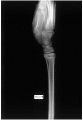 Figure 60.1 Lateral view of the right wrist demonstrates a growth plate injury. Note that the physis is not even across its entire width; it is wider at the dorsal aspect and narrower on the ventral side. This pattern signifies a Salter-Harris type I fracture. |
Avulsion Fractures
Avulsion fractures, although not characteristic of children, occur when a fragment is pulled off a bone by an attached tendon or muscle. A common site is at the base of the fingers’ phalanges or at the pelvic iliac crest. They are best seen on the lateral radiographic view.
COMMON CLINICAL ENTITIES
Clavicle Fractures
Fractures of the clavicle occur when a child lands directly on the lateral aspect of a shoulder, and the force is transmitted medially. Greenstick fractures predominate in the middle third of the clavicle, but fractures can occur at the distal end. They are best treated with reassurance and measures for comfort. Slings can be used for older children to avoid painful arm dangling. Local ice and oral analgesics are also indicated. Figure-of-eight wraps are not useful and may cause harm when they impinge on the axillary area. Neonates can present with a healing callus of the clavicle in the first month of life after a difficult delivery and shoulder dystocia. Radiographic confirmation is necessary only to allay parental fears.
Nursemaid’s Elbow
“Nursemaid’s elbow,” or more accurately dislocation or tearing of the annular ligament from the radial head, is a common entity in children between 4 months and 8 years of age; the peak incidence is from 15 to 30 months. Dislocation or subluxation of the elbow does not occur; the articular cartilages remain in direct proximity. This injury is more common in girls than boys and more common in the left than the right arm. Although a variety of histories can be elicited, a “pulling” theme is evident in all cases.
Stay updated, free articles. Join our Telegram channel

Full access? Get Clinical Tree


