Microcephaly
Susan I. Blaser, MD, FRCPC
DIFFERENTIAL DIAGNOSIS
Common
Secondary/Acquired from
Hypoxic Ischemic Encephalopathy
TORCH Infections
Nonaccidental Trauma
Meningitis
Fetal Alcohol Syndrome
Less Common
Primary/Genetic with
Gyral Simplification
Cortical Dysplasia
Midline Anomaly
Cerebellar Hypoplasia
Hypomyelination
Rare but Important
Microlissencephaly
Pseudo-TORCH
Aicardi-Goutières
Progeroid Syndromes
Cockayne
ESSENTIAL INFORMATION
Key Differential Diagnosis Issues
Was head circumference ever normal?
Decreased cranio-facial ratio on sagittal view helpful, tape measure best
Do not disregard nonaccidental trauma as cause of small head
Helpful Clues for Common Diagnoses
Hypoxic Ischemic Encephalopathy
Perinatal or birth asphyxia (asphyxia neonatorium)
Patterns helpful, even if no history
Profound: Atrophy, gliosis
Posterior putamen
Lateral thalami
Rolandic cortex
Prolonged progressive
Typical watershed encephalomalacia
Mixed
Features of both, ± calcified thalami
TORCH Infections
Cytomegalovirus (CMV) (common) and rubella (rare, today) most likely to have microcephaly
Toxo, CMV, HIV, and rubella may have intracranial Ca++, which may help in determining underlying etiology
CMV: Parenchymal or periventricular Ca++
HIV: Basal ganglia and subcortical calcifications
Toxo: Parenchymal or periventricular Ca++
Rubella and HSV cause lobar brain destruction/encephalomalacia
CMV may also have cortical dysplasia, periventricular Ca++, hypomyelination
Nonaccidental Trauma
History is crucial
Majority of abuse in children < 1 year old
Most common in children 2-6 months old
After trauma: Subarachnoid and subdural hemorrhages are common
Subdural hemorrhage over convexity, interhemispheric, overlying tentorium
Caution in attempting to determine age of subdural hemorrhage
BUT look for evidence of trauma/fractures on ALL available films
Brain imaging of microcephaly
Global atrophy or hemiatrophy
Hemosiderin
Meningitis
Early infancy: Group B strep most damaging
Hypothalamus
Chiasm
Inferior basal ganglia
Diffuse cortex, often asymmetric
Fetal Alcohol Syndrome
Microcephaly
By tape measure
Or MR volumetrics
Anomalies may occur, but not specific
Diffusion tensor imaging (DTI) reported to show abnormal connectivity
Helpful Clues for Less Common Diagnoses
Gyral Simplification
Small, grossly normal brain
Looks like “small but perfect” brain
Corpus callosum may appear thick, lack isthmus
Cortical Dysplasia
Midline Anomaly
Holoprosencephaly
Most severe are smallest
Agenesis of corpus callosum
Assess corpus callosum presence, size, shape
Is there isthmus?
Cerebellar Hypoplasia
In multiple syndromes
Olivopontocerebellar degeneration
Spinocerebellar ataxia
Carbohydrate deficient glycoprotein syndrome type 1a
May be clue to rare disorders
Microlissencephaly
TUBA1A mutations: Lissencephaly PLUS cerebellar hypoplasia
Assess degree of deficiency
Fastigial recess, primary fissure
Degree of vermian lobulation
Tegmento-vermian angle (is the inferior 4th ventricle open?)
Hypomyelination
May be a clue to rare disorders
Early onset West syndrome with cerebral hypomyelination and reduced white matter
3-phosphoglycerate dehydrogenase deficiency
Progressive encephalopathy, edema, hypsarrhythmia, optic atrophy (PEHO)
Helpful Clues for Rare Diagnoses
Microlissencephaly
Z-shaped brainstem
Callosal agenesis
Surface often totally smooth
Very small brain
Pseudo-TORCH
Aicardi-Goutières
CMV-like: Ca++
Hypomyelinaton
Atrophy
Autosomal recessive, important to diagnose
Elevated CSF α-interferon
Early onset: TREX1 mutation
Late onset: RNASEH2B mutation
Progeroid Syndromes
Cockayne
Cachectic dwarfism with mental retardation
Disorder of DNA repair: Several mutations known, but lack phenotype-genotype correlation
Facies & neuroimaging progressive
Basal ganglia/dentate Ca++
Demyelination
Atrophy
Image Gallery
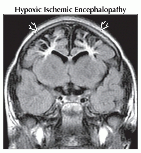 Coronal FLAIR MR shows cystic encephalomalacia  in the border zone distribution in this 3 year old with a history of peripartum prolonged partial asphyxia. in the border zone distribution in this 3 year old with a history of peripartum prolonged partial asphyxia. |
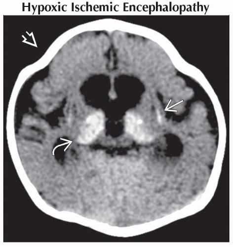 Axial NECT in a 3-month-old infant shows fusion of the coronal sutures
 due to severe brain volume loss, shrunken and calcified putamina due to severe brain volume loss, shrunken and calcified putamina  and thalami and thalami  following severe mixed HIE. following severe mixed HIE.Stay updated, free articles. Join our Telegram channel
Full access? Get Clinical Tree
 Get Clinical Tree app for offline access

|