Fig. 27.1
A clinical photograph showing a newborn with abdominal distension secondary to meconium ileus
Meconium ileus is divided into simple or complicated.
Simple meconium ileus : There is simple obstruction of the terminal ileum. The distal small bowel (terminal ileum) contains inspissated, thick viscous meconium, and proximal to this the ileum is distended while the colon is small and unused (microcolon).
Complicated meconium ileus : can be associated with:
Volvulus
Bowel gangrene
Perforation
Intestinal atresia
Pseudocyst formation
Perforation
Meconium peritonitis
Volvulus occurs due to the distended loops of terminal ileum and can lead to vascular compromise and bowel gangrene.
Perforation can occur and causes meconium peritonitis. This is generally a sterile, chemical peritonitis that causes irritation of the peritoneal surfaces. With healing, calcifications can take place.
Differential Diagnosis
Meconium plug syndrome, ileal atresia, colonic atresia, intestinal pseudo-obstruction, and Hirschsprung’s disease
Investigations
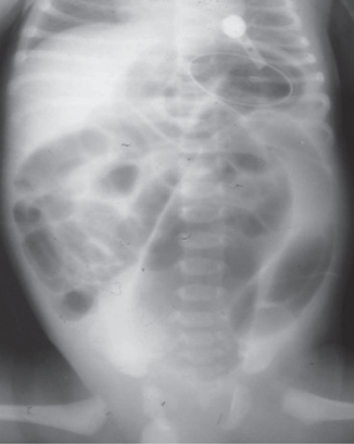
Plain abdominal radiographs : These are usually nonspecific and show multiple dilated loops of small bowel. The thick meconium has a ground-glass appearance and when mixed with air gives the intestinal contents a granular soap-bubble appearance (Fig. 27.2).

Fig. 27.2
Plain abdominal radiograph showing dilated bowel loops
The soap-bubble appearance may be seen also in Hirschsprung’s disease, ileal or colonic atresia, or meconium plug syndrome.
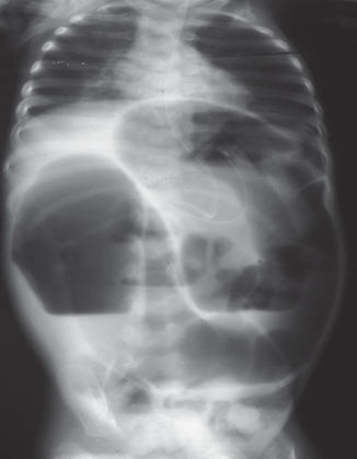
Air-fluid levels may be seen in the dilated small bowel. The presence of air-fluid levels should raise the possibility of associated intestinal atresia (Fig. 27.3).

Fig. 27.3
Abdominal radiograph showing dilated bowel loops with air-fluid levels
Stay updated, free articles. Join our Telegram channel

Full access? Get Clinical Tree


