Fig. 26.1
Diagrammatic representation of Meckel’s diverticulum. It is a true diverticulum composed of all the three layers and lies on the antimesenteric border of the intestines
It is present in approximately 2 % of the population.
It was first described by Fabricius Hildanus in the sixteenth century and later named after Johann Friedrich Meckel, who described its embryological origin in 1809.
Meckel’s diverticulum may harbor abnormal tissues including jejunum, duodenal mucosa, or Brunner glands (2 % of ectopic cases).
Heterotopic rests of gastric mucosa and pancreatic tissue are seen in 60 and 6 % of cases respectively.
The prevalence of Meckel’s diverticulum in males is three to five times higher than in females.
The majority of Meckel’s diverticulum are asymptomatic and only 2 % of cases are symptomatic.
Meckel’s diverticulum is located in the distal ileum, usually within about 60–100 cm (2 ft) of the ileocecal valve.
It is typically 3–5 cm long, runs antimesenterically, and has its own blood supply.
The rule of 2s for Meckel’s diverticulum:
2 % of the population.
2 ft from the ileocecal valve.
2 in. (in length).
2 % are symptomatic.
2 types of common ectopic tissue (gastric and pancreatic).
2 years is the most common age at clinical presentation.
2 times more boys are affected.
Meckel’s diverticulum can also present in an indirect hernia , typically on the right side, where it is known as a “Littré Hernia.”
Furthermore, it can be attached to the umbilical region by the vitelline ligament, with the possibility of vitelline cysts, or even a patent vitelline canal forming a vitelline fistula when the umbilical cord is cut. Torsion of intestine around the intestinal stalk may also occur, leading to obstruction, ischemia, and necrosis.
Embryology
Embryologically , the omphalomesenteric duct (omphaloenteric duct, vitelline duct, or yolk stalk) connects the embryonic midgut to the yolk sac ventrally, providing nutrients to the midgut during embryonic development.
Subsequently, the vitelline duct narrows progressively and disappears between the 5th and 8th weeks of gestation.
Sometimes, the proximal part of vitelline duct fails to regress and involute, and remains as a remnant of variable length forming Meckel’s diverticulum (Fig. 26.2).
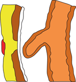
Fig. 26.2
Diagrammatic representation of Meckel’s diverticulum
Meckel’s diverticulum lies on the antimesenteric border of the ileum and extends into the umbilical cord of the embryo.
The left and right vitelline arteries originate from the primitiv e dorsal aorta, and travel with the omphaloenteric duct. The right branch becomes the superior mesenteric artery that supplies a terminal branch to Meckel’s diverticulum, while the left involutes.
Other possible omphaloenteric duct anomalies include:
An omphalomesentric ligament/fibrous band (Fig. 26.3).
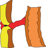
Fig. 26.3
Diagrammatic representation of an omphalomesenteric fibrous band attached to the site of Meckel’s diverticulum on the intestines side and site of umbilicus
An omphalomesenteric fistula (Fig. 26.4).
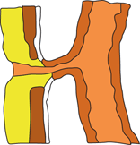
Fig. 26.4
Diagrammatic representation of an omphalomesenteric fistula. Note the communication between the intestines and umbilicus
An omphalomesentric cyst (Fig. 26.5).
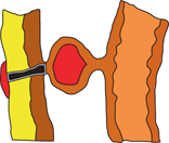
Fig. 26.5
Diagrammatic representation of an omphalomesenteric cyst. The cyst is attached to the site of Meckel’s diverticulum on one side and the umbilicus on the other side
A persistent vitelline artery running along the fibrous cord which connects the ileum to the umbilicus.
An umbilical sinus (Fig. 26.6).
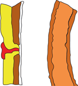
Fig. 26.6
Diagrammatic representation of an umbilical sinus
Meckel’s diverticulum may also contain heterotropic tissues as follows:
Gastric mucosa (60 %).
Pancreatic tissue (6 %).
Both pancreatic tissue and gastric mucosa (5 %).
Jejunal mucosa (2 %).
Brunner tissue (2 %).
Both gastric and duodenal mucosa (2 %).
Rarely, colonic, rectal, endometrial, and hepatobiliary tissues have been noted.
Symptoms
The majority of people with Meckel’s diverticulum are asymptomatic.
Stay updated, free articles. Join our Telegram channel

Full access? Get Clinical Tree


