Type I: Tumor at diaphyseal location or metaphyseal location over 2 cm from the epiphyseal plate
Type II: Tumor at metaphyseal location extending to less than half of the epiphyseal growth plate
Type III: Tumor at metaphyseal location extending to the whole growth plate
Type IV: Metaphyseal tumor extending through the growth plate into part of the epiphysis at least 10 mm from the articular surface
Type V: Tumor extending into less than half of the epiphysis less than 1 cm away from the articular surface
Type VI: Tumor extending into more than half of the epiphysis less than 1 cm away from the articular surface
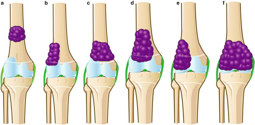
Fig. 21.1
Classification of tumor excision. (a) Type I. (b) Type II. (c) Type III. (d) Type IV. (e) Type V. (f) Type VI
Surgical Techniques
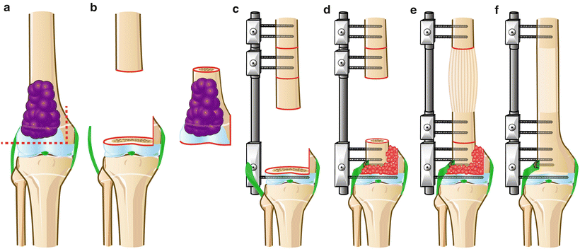
Type I: DO Method (Fig. 21.2)
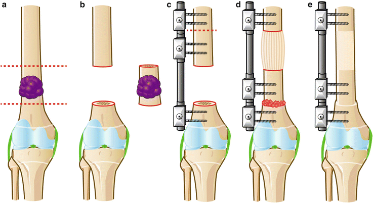
Fig. 21.2
Type I: DO method. (a) Resection line. (b) Tumor resection. (c) Application of external fixator and osteotomy for lengthening. (d) Bone grafting at docking site. (e) After bone consolidation
In Type I, the tumor is diaphyseally or metaphyseally located over 2 cm away from the epiphyseal plate. The operative technique of the DO method consists of en bloc tumor excision with preservation of the epiphyseal plate and articular surface. An external fixator is applied and osteotomy is performed for postoperative bone transport. After docking bone transport, we usually perform bone graft at the docking site. Shortening-distraction is indicated if acute shortening is possible during the operation. Optional intramedullary nailing, if feasible, or plate conversion, is also used to shorten the external fixation time [4, 6].
Type I: LN Method (Fig. 21.3)
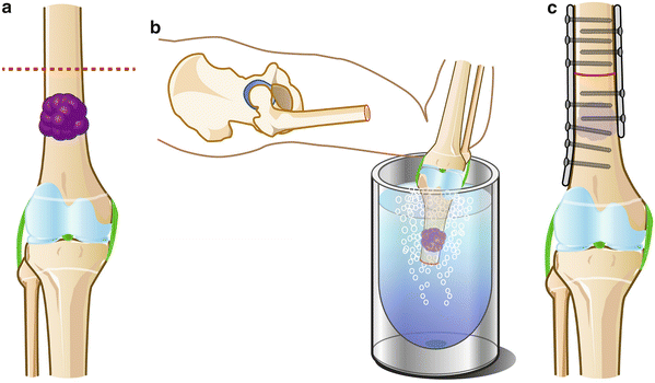
Fig. 21.3
Type I: LN method. (a) Resection line. (b) Pedicle freezing. (c) Reconstruction with double plates
One-site osteotomy and pedicle freezing method are applied in the Type I LN method. After transdiaphyseal osteotomy, soft tissue is removed, and the tumor is curetted after thorough isolation to prevent fracture during freezing. Bony lesions connected to the limb are then rotated and frozen in liquid nitrogen for 20 min, thawed at room temperature for 15 min, and then thawed in distilled water for 10 min. Reconstructions are performed by osteosynthesis using double plates [8].
Type II: DO Method (Fig. 21.4)
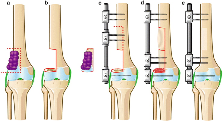
Fig. 21.4
Type II: DO method. (a) Resection line. (b) Tumor resection. (c) Application of external fixator and osteotomy for lengthening. (d) Bone grafting at docking site. (e) After bone consolidation
In Type II, the tumor is located metaphyseally and extends to less than half of the epiphyseal growth plate. There is also a possibility of joint deformity due to unequal growth after reconstruction. The operative technique of the DO method consists of hemicortical en bloc tumor excision. An external fixator is applied, and proximal hemicortical osteotomy is performed for postoperative bone transport. Epiphysiodesis of the other side of the growth plate is one option to avoid future angular deformities for young patients. After docking bone transport, we usually perform a bone graft at the docking site. At that time, plate conversion is optionally used to shorten the external fixation time. If a deformity occurs in the future, correction with or without lengthening is performed.
Type II: LN Method (Fig. 21.5)
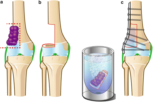
Fig. 21.5
Type II: LN method. (a) Resection line. (b) Freezing tumor-bearing bone. (c) Reconstruction with single plate
Hemicortical resection and a free-freezing method are applied for Type II resections. After transepiphyseal and hemicortical resection, soft tissue is removed, and the tumor is curetted after thorough isolation. The tumor-bearing bone is frozen in liquid nitrogen and thawed as usual. Reconstruction is performed by osteosynthesis using a single-locking plate [7].
Type III: DO Method (Fig. 21.6)
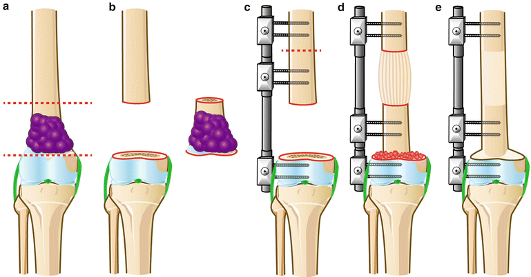
Fig. 21.6
Type III: DO method. (a) Resection line. (b) Tumor resection. (c) Application of external fixator and osteotomy for lengthening. (d) Bone grafting at docking site. (e) After bone consolidation
In Type III, the tumor is metaphyseally located and extends to the whole growth plate. Limb length discrepancies are anticipated in immature patients, which may be managed surgically if necessary. The operative technique of the DO method consists of transepiphyseal and intercalary en bloc tumor excision with the growth plate. Surgical technique is basically same as in the Type I DO method.
Type III: LN Method (Fig. 21.7)
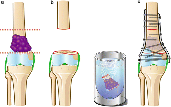
Fig. 21.7
Type III: LN method. (a) Resection line. (b) Freezing tumor-bearing bone. (c) Reconstruction with double plates
Intercalary resection and the free-freezing method are applied for Type III resection. After transepiphyseal and intercalary resection, free freezing is performed in the same way as in the Type II LN method. Reconstruction is performed by osteosynthesis using double plates.
Type IV: DO Method (Fig. 21.8)