Malignancies of the Vulva


Malignancies of the Vulva


Mitchel S. Hoffman

Xiaomang B. Stickles


INTRODUCTION

Carcinoma of the vulva is an uncommon malignancy accounting for only 2% of all female genital malignancies. It is predominantly a disease of older women. In this country as a whole, the steady increase in life expectancy has brought carcinoma of the vulva into a place of more importance among gynecologic malignancies. The predominant histologic type is squamous cell carcinoma, which accounts for about 90% of the tumors in most series. This malignancy metastasizes primarily through the lymphatic system in an orderly manner through the superficial inguinal, deep inguinal, and pelvic lymph nodes.

During the first half of the 20th century, the absolute 5-year survival rate for carcinoma of the vulva was 15%. During the late 19th and early 20th centuries, principles of surgical oncology were developed and put into clinical practice. The application of these surgical principles to the treatment of vulvar cancer resulted in a significant improvement in survival. The survival of patients with invasive squamous cell carcinoma of the vulva is dependent on a number of histopathologic factors, but it most closely relates to the pathologic status of the inguinal lymph nodes. Until the early 1980s, patients with invasive carcinoma of the vulva were routinely treated with radical vulvectomy and bilateral inguinofemoral and pelvic lymphadenectomy. Accumulation of data on vulvar cancer during the mid- to late 20th century led to earlier diagnosis and a better understanding of the nature and modes of spread of this disease. Especially over the past several decades, treatment of this malignancy has undergone a number of significant modifications resulting in less radical surgery with good survival rates and significantly better quality of life for women with this malignancy.

This chapter first reviews the epidemiology, clinical characteristics, staging, and prognostic factors for invasive carcinoma of the vulva. The broad spectrum of treatment of this disease is then reviewed with an emphasis on the surgical treatment. Finally, histologic variants of vulvar malignancy and their treatment are discussed individually.


EPIDEMIOLOGY

Invasive squamous cell carcinoma of the vulva is typically a disease of postmenopausal women, with a median age at diagnosis of about 65 years (Fig. 49.1). However, the age range is wide, and there appears to be an increasing incidence in younger women. This has been mainly attributed to infection with the human papillomavirus (HPV), which accounts for approximately 60% of vulvar squamous cell carcinoma. HPV subtypes 18 and 33 account for about half of HPV-associated vulvar cancers. HPV does not appear to play as significant of a role in the pathogenesis of invasive squamous cell carcinoma of the vulva in older women, who frequently have coexisting lichen sclerosis. In the older population, vulvar carcinoma commonly arises from a background of chronic inflammation without evidence of HPV involvement. Other sexually transmitted factors that have been epidemiologically associated with vulvar cancer are the granulomatous venereal diseases, especially in countries where these are prevalent.

It is to yet to be determined what effect the implementation of HPV vaccination will have on the incidence of vulvar squamous cell carcinoma. The full impact will likely not be known for many decades as vulvar carcinoma is most common in older women.

Vulvar carcinoma in situ (Fig. 49.2), like cervical carcinoma in situ, is considered a precursor to invasive disease, although the risk of progression appears to be lower. Although Jones and Rowan found in 7 of 8 women with untreated vulvar intraepithelial neoplasia (VIN) that invasive cancer developed within 8 years, three other studies found progression rates of 5% to 16%. In a recent systematic review of 3,322 published patients, 8 of 88 untreated women with VIN 3 progressed to invasive cancer in 12 to 96 months. In a large review, 3.2% of women treated with partial or total vulvectomy for VIN were found to have occult invasive cancer. However, several selected series showed higher rates of 16% to 22%. The risk
for invasive cancer may be greater with perianal location, increased age, immunosuppression, and previous radiotherapy.


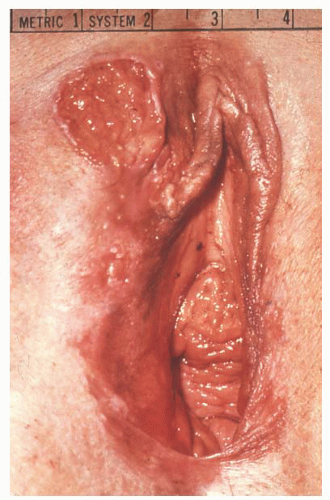
FIGURE 49.1 Invasive squamous cell carcinoma of the vulva. This is a 1.8-cm ulcerative lesion on the right anterior vulva.

Vulvar carcinoma in situ tends to be multifocal with a lower risk of invasive cancer in younger women, but it tends to be unifocal with a higher risk of invasive disease in older women. Most patients with VIN 3 should be treated, and long-term follow-up is mandatory. Although progression of VIN from grade 1 to grades 2 and 3 has been demonstrated, in the absence of atypical changes, other vulvar epithelial abnormalities do not appear to have significant precancerous potential. Patients who have cervical neoplasia are at increased risk for developing vulvar neoplasia and vice versa. This so-called field phenomenon should heighten the physician’s surveillance for the development of other lesions once a lower genital tract neoplasm occurs.


FIGURE 49.2 Carcinoma in situ of the vulva. Vulvar intraepithelial lesions can be white, brown, or red.

Hypertension and diabetes mellitus are common in patients with invasive vulvar cancer, but this may simply be related to the elderly population affected. The association of vulvar cancer with obesity is unclear. There does not appear to be any significant association with parity or race. Smoking is associated with an increased risk of vulvar cancer. One group that does appear to be at particular increased risk for the development of invasive vulvar cancer is chronically immunosuppressed women.


CLINICAL PRESENTATION

The most common initial symptom of vulvar cancer is pruritus, which may be of long duration. Vulvar pain, discharge, and bleeding are less commonly reported. The patient often becomes aware of a lesion on her vulva; but because of the superficial nature of the lesion, delay in seeking medical help is common. Physician delay has also been commonly reported, often related to prolonged empiric medical treatment before a biopsy is done. Invasive squamous cell carcinoma of the vulva may present as an exophytic, cauliflower-like lesion or it may have an ulcerative appearance (Fig. 49.3). It involves the labia majora in about two thirds of patients. The remaining tumors involve the clitoris, labia minora, posterior fourchette, and perineum. These cancers can be exophytic, ulcerating, or flat. Rarely, a patient may present with erosive tumor growth from the groin nodes if symptoms have been ignored for long periods of time.


FIGURE 49.3 Invasive carcinoma of the left vulva with extensive associated carcinoma in situ involving both labia.


HISTOLOGY

Squamous cell carcinoma accounts for about 90% of the invasive vulvar malignancies. Melanoma is the second most common histologic type, accounting for 5% to 10% of vulvar cancers. Some of the less common vulvar malignancies include Bartholin gland carcinoma (either adenocarcinoma or squamous carcinoma), basal cell carcinoma, verrucous carcinoma, invasive Paget disease, and sarcomas.


PRETREATMENT INVESTIGATION


Biopsy

The relative rarity of vulvar cancer and the general lack of awareness of typical signs and symptoms, even by medical professionals, frequently lead to a delay in diagnosis. These observations underscore the need for patient and physician education with regard to the early diagnosis of carcinoma of the vulva. Therefore, biopsy of any questionable vulvar lesion prior to empiric treatment is warranted. Vulvar biopsy is a simple procedure that can be done in the office under local anesthesia. Biopsy technique is demonstrated in Chapter 23 (Fig. 23.1).


Preoperative Clinical Evaluation

Patients with carcinoma of the vulva are generally elderly and often have coexisting medical problems. A thorough preoperative evaluation by internal medicine and anesthesiology consultants should be requested when appropriate. As previously discussed, these women have an increased incidence of cervical and vaginal neoplasia, indicating the importance of a careful pelvic examination and Pap test screening.


Imaging Studies

Beyond physical examination and a chest radiograph, routine studies to rule out metastases are generally not indicated except in the presence of locally advanced disease. Cystoscopy, intravenous pyelography, or proctoscopy (or all three) is indicated if it appears that locally advanced cancer may involve the bladder, urethra, or rectum. Magnetic resonance imaging (MRI) or computed tomography (CT) may help to determine resectability and treatment planning. When a locally advanced tumor or inguinal lymph node enlargement is noted, CT scan of the groins, pelvis, and abdomen is suggested. This aids in determining the resectability of the tumor and metastases, extension to the pelvic lymph nodes, and distant metastases (Fig. 49.4). Increasingly, positron emission tomography (PET) has been utilized to evaluate the presence or absence of metastatic disease, though scant data exist to support its use in vulvar cancer (Fig. 49.5). In a small series of 15 patients reported by Cohn and colleagues, the sensitivity of PET in detection of pathologically confirmed groin metastasis was 67% and specificity was 95%. Another recent analysis by Kamran et al. showed a sensitivity of only 50% and specificity and positive predictive value of 100%. Larger studies validating its utility are lacking. Due to limited sensitivity, omission of lymphadenectomy should not be based on PET results alone.


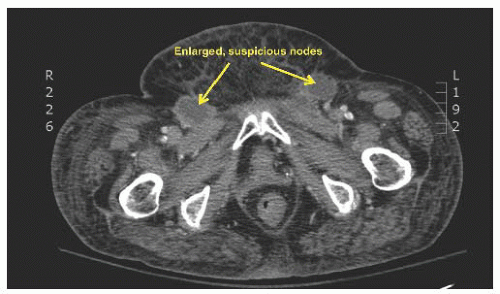
FIGURE 49.4 Computerized tomography (CT) image of the inguinal area showing enlarged lymph nodes typical for metastatic cancer (arrows).


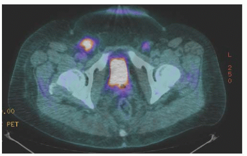
FIGURE 49.5 Positron emission tomography (PET) of the groin areas showing significantly increased FGD uptake in bilateral nodes very typical of metastatic cancer (same patient as in Fig. 49.4).


STAGING


Route of Spread

Squamous cell carcinoma of the vulva can spread by local extension to involve the vagina, urethra, anus, or pubic bone. Metastasis is primarily via the lymphatic system, but hematologic spread is also possible. The lymphatics of the labia communicate to the inguinal lymph nodes. The lymphatics of the perianal area drain in a similar manner, but lesions that involve the vagina, anus, or rectovaginal septum can drain directly into the pelvic lymph nodes. Although there are channels that interconnect the clitoris to the deep pelvic nodes, it appears that they are of minimal clinical significance. The lymphatics of the vulva are numerous and tend to cross the midline. The regional lymph nodes include the superficial inguinal nodes, the deep inguinal-femoral nodes, and the pelvic lymph nodes. The superficial inguinal lymph nodes are the primary nodal group of the vulva. Depending on the location of the primary carcinoma, one or two of these superficial inguinal nodes is the first site of metastatic spread. This node is referred to as the sentinel lymph node. Because of the predictably orderly progression of lymph node metastases in vulvar cancer, involvement of the deeper femoral lymph nodes or the pelvic nodes is very infrequent if the sentinel node(s) is uninvolved. These superficial inguinal lymph nodes drain through the cribriform fascia to the femoral lymph nodes. Drainage from here is under the inguinal ligament into the pelvic lymph nodes. The pelvic lymph nodes are virtually never positive in the absence of inguinofemoral lymph node metastases. The invasion of lymph node metastases roughly correlates with the size of the primary tumor.


TABLE 49.1 2009 FIGO Staging System for Vulvar Cancer


STAGE


EXTENT


IA


Lesion ≤2 cm and stromal invasion ≤1 mm, confined to vulva or perineum, no nodal metastasis


IB


Lesion >2 cm or stromal invasion >1 mm, confined to vulva or perineum, no nodal metastasis


II


Tumor of any size with extension to adjacent perineal structures (one-third lower urethra, vagina, or anus), no nodal metastasis


III


Tumor of any size with or without extension to adjacent perineal structures with positive inguinofemoral lymph nodes


IIIA


(i) With one lymph node metastasis (≥5 mm)


(ii) One to two lymph node metastasis (<5 mm)


IIIB


(i) With two or more lymph node metastasis (≥5 mm)


(ii) With three or more lymph node metastasis (<5 mm)


IIIC


With positive nodes with extracapsular spread


IVA


(i) Upper urethral and/or vaginal mucosa, bladder mucosa, rectal mucosa, or fixed to pelvic bone


(ii) Fixed or ulcerated inguinofemoral lymph nodes


IVB


Distant metastasis, including pelvic lymph nodes


FIGO Staging System

In 1979, the International Federation of Gynecology and Obstetrics (FIGO) approved a clinical classification for invasive squamous cell carcinoma of the vulva. This was based on an analysis of tumor (T) by size and location; node (N) status by palpation; distant metastases (M) as assessed by physical examination; endoscopic evaluation of the bladder or rectum, or both; and radiologic investigation. Most patients with invasive carcinoma of the vulva are treated surgically, and it was recognized that the clinical evaluation of inguinal lymph node metastases was often inaccurate. Therefore, in 1988, FIGO approved a surgical staging system. This staging system has undergone several revisions over the years as new surgical pathologic findings have been correlated with outcome. The most recent FIGO staging classification adopted in 2009 is shown in Table 49.1.

Stage I is divided into stages IA and IB because primary tumors less than 2 cm in diameter with very superficial invasion have a very low incidence of lymph node involvement and these patients have an excellent prognosis. However, a number of patients with small tumors have been reported with metastatic disease, which emphasizes the importance of surgical staging whenever possible (Table 49.2).

In the FIGO staging revision of 2009, the involvement of lower one third of the urethra, vagina, or anus was downgraded from stage III to stage II. Stage III was subdivided to reflect the prognostic significance of both the number and extent of lymph node involvement.

With increasing use of sentinel lymph node biopsy for staging and improvements in anesthetic techniques and perioperative care, it is rare today that a patient cannot be surgically staged, but the old clinical staging system should be used with appropriate notation in such patients.


TABLE 49.2 Tumors with Invasion of Less Than 1 mm with Nodal Disease, Either at Diagnosis or Recurrence


INVESTIGATOR


PATIENT AGE


TUMOR SIZE (CM)


TUMOR GRADE


TUMOR DEPTH (MM)


CAPILLARY OR LYMPHATIC SPACE INVOLVEMENT


MONTHS TO DIAGNOSIS OF NODAL DISEASE


STATUS


Sedlis (1987)


61


2.5


4a


1


0


NA


Atamdede (1989)


75


1


1


0.72


No


13


AWD


van der Valden (1992)


84


1


3


0.3


No


20


DOD


Kelley (1992)


52


NA


NA


<1


NA


31


A at 27 mo


Stehman (1992)


57


2


3


0.6


NA


26


A at 35 mo


Thangavelu (2006)


64


NA


2


<1


No


3


DOD


51


NA


1


0.9


No


36


DOD


Volgger (1997)


39


0.3


1


<1


No


4


DOD


Iyibozkurt (2010)


62


2


NA


<1


NA


27


DOD


a GOG grading system.


NA, not available; AWD, alive with disease; DOD, dead of disease; A, alive.


PROGNOSTIC FACTORS


Primary Tumor Characteristics

Primary tumor factors that appear to have prognostic importance include tumor diameter, depth of invasion or tumor thickness, tumor differentiation, lymph-vascular space involvement, and surgical margin status. Tumor involvement of the distal urethra, vagina, or perineum is also an adverse prognostic factor. Sedlis and colleagues reported on a Gynecologic Oncology Group (GOG) study of 272 patients with early vulvar cancer less than or equal to 5 mm in tumor thickness. They found that histologic grade, capillary-like space involvement, tumor thickness, and clitoral or perineal location were all significant predictors of groin node metastases. A subsequent GOG study reported by Homesley and associates on 588 evaluable patients with invasive carcinoma found lymph-vascular space involvement, tumor differentiation, age, and tumor thickness to be significant independent risk factors for groin node metastases. Factors that predicted an increased risk of local recurrence included margin status, large tumor size, and deep invasion. Tumor extending to less than 8 mm from surgical margin was associated with an increased risk of recurrence.

A few histologic variants of squamous cell carcinoma have been associated with a more favorable prognosis, including verrucous carcinoma (discussed later), warty carcinoma, carcinoma in situ with early stromal invasion, and keratoacanthoma.


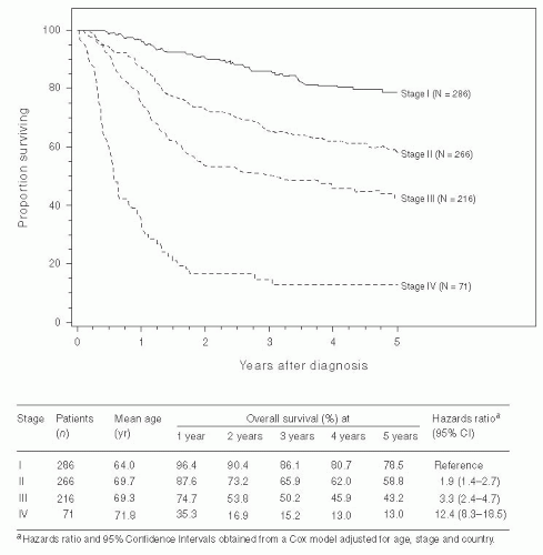
FIGURE 49.6 Survival curves for patients with squamous cell carcinoma of the vulva related to FIGO stage. These patients were treated from 1999 through 2001 and were staged according to the FIGO staging system of 1996. (Reprinted from Beller U, Benedet JL, Creasman WT, et al. Carcinoma of the vagina: 26th Annual report on the results of treatment in gynecological cancer. Int J Gynecol Obstet 2006;95:S29-S42, with permission. Copyright © 2006 International Federation of Gynecology and Obstetrics. Published by Elsevier Ireland Ltd. All rights reserved.)


Lymph Node Status

The status of the groin nodes is clearly the most important prognostic factor for patients with invasive squamous cell carcinoma of the vulva. This disease has a relatively low propensity for distant metastases. Recurrences tend to be local or regional, and even unremitting disease tends to remain locoregional for long periods of time.

The overall 5-year survival rate for all treated patients is about 60%, with a corrected 5-year survival rate of about 70%. In the GOG study previously mentioned, 65.5% of the patients had negative groin nodes and 34.5% had positive nodes; this is consistent with other reports in the literature. In this large surgically treated series, the survival rate was 90.9% for patients with negative lymph nodes and 57.7% for patients with lymph node metastasis. When the pelvic lymph nodes are known to be positive, the survival rate decreases to about 20%. Figure 49.6 shows the survival of patients with squamous cell carcinoma of the vulva staged with the older FIGO staging classification. No large studies have reported long-term results with the 2009 staging.

Metastatic involvement of multiple lymph nodes further decrease the prognosis that was recognized in the 2009 FIGO staging system, which subdivided stage III disease depending on the number of nodes involved and the size of the metastatic deposits. Several studies have shown that when only one lymph node is involved, survival is still quite good. However, survival decreases drastically with metastases to three or more
nodes or with bilateral nodal involvement. On the basis of limited data from a few studies, it appears that large nodal diameter, extensive nodal replacement, and especially extracapsular extension of a lymph node metastasis are adverse prognostic factors. In a report by Origoni and colleagues in 1992 that was based on 53 vulvar cancer patients with groin node metastases, the survival rate varied from 90.9% when the diameter of the metastasis was less than 5 mm to 20% when it was larger than 15 mm, and from 85.7% to 25% when the metastases were intracapsular and extracapsular, respectively. Especially important are the data from the 19 patients in that study with a single positive node. For these patients, the 5-year cancerrelated survival rate was 90% when the metastasis diameter was less than 5 mm versus 37.5% when it was 15 mm, and 85.7% when the metastasis was intracapsular versus 20% when it was extracapsular. Results from a study by Paladini and associates were similar; they reported that patients with intracapsular metastases tended to have recurrence at distant sites, whereas patients with extracapsular nodal disease were more likely to have local or groin recurrence.


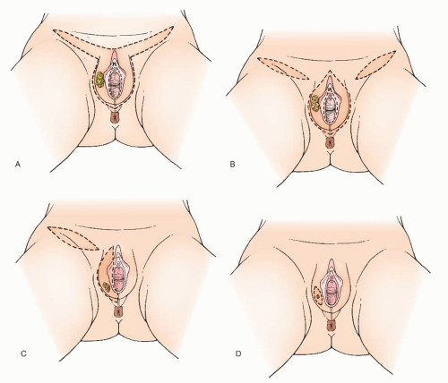
FIGURE 49.7 Modification in regional lymph node management. A: En bloc removal. B: Radical vulvectomy with bilateral inguinal-femoral lymphadenectomy through three separate incisions. C: Unilateral lymphadenectomy for a well-lateralized lesion. D: Early lesion with omission of lymphadenectomy.


TREATMENT OF INVASIVE SQUAMOUS CELL CARCINOMA OF THE VULVA


Historical Perspective

In the 1940s, Taussig and Way reported favorable outcomes in carcinoma of the vulva after radical vulvectomy with bilateral inguinal lymphadenectomy performed by en bloc excision. This radical procedure then became the standard therapy applied to most patients. It involved radical removal of the entire vulva, the mons pubis, the inguinofemoral lymph nodes, and often the pelvic lymph nodes. The large surgical defect thus created was generally closed under tension with a high subsequent breakdown rate, significant postoperative morbidity, and marked disfigurement of the genital area. Important concerns with this approach for the treatment of vulvar cancer led to a number of modifications, especially over the past 25 years (Fig. 49.7). Some of these concerns include the high rate and the severity of wound complications and the psychosexual effects of radical removal of the vulvar tissues. Other potential problems related
to the en bloc radical resection include urinary or fecal incontinence and vaginal prolapse. The “one-size-fits-all” approach of treating all patients with the same radical vulvectomy and inguinal-femoral lymphadenectomy resulted in overtreatment of many patients and undertreatment of some with inadequate attention to local margin and the risk of lymph node metastasis. The following section provides a more individualized approach based on current literature and extensive surgical experience.


Primary Surgery: Selection of Appropriate Primary Operation


Appropriate Candidates for Primary Surgery

Certain tumor characteristics may preclude an otherwise medically fit patient from undergoing primary surgery. Proximity to important functional structures such as the urethra, clitoris, and anal sphincter must be considered. If adequate surgical margins cannot be obtained without sacrificing such a structure, neoadjuvant treatment with radiation and/or chemotherapy should be considered. Very large primary tumors may leave a large defect after primary resection, which may prove challenging to close. These patients may be candidates for neoadjuvant radiation plus chemotherapy to shrink the size of the primary cancer before surgical resection. The patient’s ability to tolerate a radical resection must also be taken into consideration. Some elderly patients or those with significant comorbidities who may not recover from a radical resection would tolerate a more limited resection after neoadjuvant treatment.


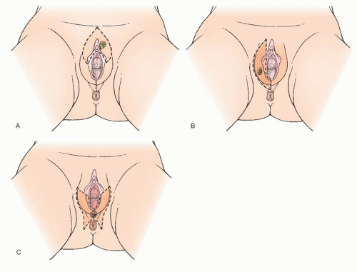
FIGURE 49.8 Modified radical vulvectomy. A: Anterior hemivulvectomy. B: Right hemivulvectomy with clitoral sparing. C: Posterior hemivulvectomy.


Types of Incisions

The most important modification to the classic en bloc excision came after Hacker and colleagues and DiSaia and associates reported that separation of the vulvar and groin incisions resulted in a significant reduction in wound morbidity (Fig. 49.7B). The report by Hacker and colleagues in 1981 consisted of 100 patients in whom three separate incisions were used to perform the bilateral inguinofemoral lymphadenectomy and radical vulvectomy, leaving a bridge of tissue between the incisions and sparing the mons pubis. Major groin wound breakdown occurred in 14 patients, which was a considerable reduction from the 50% or higher groin wound breakdown rate generally seen with the en bloc excision. In this report, there were no isolated recurrences in either the groin or the inguinal skin bridge. There were two recurrences in the inguinal skin bridge associated with other recurrence sites; both patients originally had positive inguinal lymph nodes. In addition to these two patients, there have been other isolated reports of inguinal skin bridge recurrence. However, this still appears to be a rare event. Some authors believe that in the presence of advanced disease or grossly positive inguinal lymph nodes, an en bloc excision of the tumor and the lymph nodes is still the best approach. En bloc excision is certainly warranted at times to obtain an adequate resection of the malignancy.


Modified Radical and Radical Vulvectomy

Modified radical vulvectomy is an ambiguously defined operation that generally refers to radical removal of the portion of the vulva containing the tumor (Fig. 49.8). In recent
years, the extent of the radical vulvectomy has been modified to emphasize wide excision of the primary cancer with adequate skin and deep margins but not necessarily radically resecting the entire (uninvolved) vulva. Recommendations have included 1- to 3-cm skin margins for well-localized, unifocal lesions and to the depth of the inferior fascia of the urogenital diaphragm. The main type of morbidity relating to increasing the radicality of vulvectomy is subsequent sexual dysfunction and disfigurement. Modification of the surgical resection or the use of neoadjuvant chemoradiation should be considered to avoid compromising the function of the anus or urethra. In a 1979 study, DiSaia and colleagues reported complete preservation of sexual function in 17 of 18 patients who underwent wide local excision for early invasive tumors. They also reported that preservation of the mons pubis as well as the major portion of the superior aspect of the vulva resulted in an appreciably more satisfactory cosmetic result.

The chief concerns with decreasing the radicality of vulvectomy are the possibility of an increased risk of local recurrence and later an increased risk of a second primary vulvar cancer. Hacker and colleagues have demonstrated that the extent of the vulvectomy can be decreased as long as careful attention to the margins is observed. They compared radical vulvectomy (56 patients) with wide local excision (28 patients) in the treatment of superficially invasive squamous cell carcinoma of the vulva. The local recurrence rate was identical, 4% in both groups. A later review by Hacker and van der Velden of literature regarding patients with vulvar cancer 2 cm or less in diameter showed a local invasive recurrence rate of 7.2% for 165 patients treated with radical local excision versus 6.3% for 365 patients treated with radical vulvectomy. In a comparative study reported by Hoffman, 45 patients underwent radical vulvectomy and 45 patients were treated with modified radical vulvectomy. The local recurrence rates were 2.2% and 4.4%, respectively. Several additional studies have reported excellent local control with a modified radical vulvectomy (Table 49.3).

In a GOG study, Stehman and colleagues analyzed recurrences following modified radical hemivulvectomy. The mean time to “relapse” on the vulva was 43.4 months, and 11 of 18 patients had recurrence on the contralateral side from the primary lesion. This suggests a second cancer rather than a true local recurrence. A study by de Hullu and associates also reported a significant number of late recurrences following modified radical vulvectomy. From these results, it is apparent that women undergoing a modified vulvar operation for cancer are at increased risk for later development of a new primary vulvar tumor and require long-term close follow-up.

It appears reasonable to conclude that modified radical vulvectomy is an efficacious treatment for well-localized invasive squamous cell carcinoma of the vulva. Attention should be focused on obtaining a 2-cm gross margin around the tumor while sparing as much vulvar tissue as possible. Most patients with squamous cell carcinoma of the vulva are candidates for a modified radical vulvectomy via a separate incision from the groin lymphadenectomy. A few patients, however, by virtue of disease extent, require a traditional radical vulvectomy with radical resection of the entire vulva. Whatever the vulvar phase of the operation is called, the aim should be to excise the tumor with a 2-cm gross margin.

Because of tumor proximity, it is occasionally necessary to remove a portion of the urethra to obtain an adequate resection of a vulvar carcinoma. Although several authors have stated that removal of the outer urethra does not result in significant problems with incontinence, there are no substantial confirming data. In one small study, Reid and colleagues did find urinary incontinence to be a problem after resection of the distal urethra or even an excision close to the urethra. If a portion of the urethra is resected, a Foley catheter should be left in place (carefully taped to the leg) for about 1 week postoperatively to facilitate healing and to splint the urethra. A surgical anti-incontinence procedure should also be strongly considered at the time of resection, especially if there is any preoperative stress urinary incontinence or if more than 1 cm of the urethra has to be removed. Increasingly, consideration has been given to treating these patients with neoadjuvant radiotherapy and chemotherapy to minimize or avoid urethral resection.


TABLE 49.3 Modified Radical Vulvectomy Literature: Local Recurrence


INVESTIGATOR


NO. OF PATIENTS


LOCAL RECURRENCE (%)


MINIMUM FOLLOW-UP (MONTHS)


Di Saia (1979)


18


0


7


Hacker (1984)


28


1 (4%)


24


Burrell (1988)


28


0


16


Berman (1989)


50


4 (8%)


12


Sutton (1991)


56


7 (13%)


1


Stehman (1992)


121


10 (8%)


36


Hoffman (1992)


45


1 (2%)


12


Lin (1992)


12


2 (17%)


24


Andrews (1994)


28


2 (7%)


12


Burke (1995)


76


9 (12)


NA


de Hullu (2002)


85


7 (8%)


NA


Arvas (2005)


40


17 (43%)


NA


Total


587


60 (10%)


There is scant literature concerning the local management of vulvar cancer with perianal involvement (Fig. 49.9). However, we have noted that about one third of our patients who are referred for vulvar cancer have lesions with perianal or anal involvement. The chief problems in the management of patients with these tumors are the difficulty in obtaining adequate surgical margins while attempting to preserve anal sphincter function and deciding on which patients would be better treated either with a more radical excision and colostomy or with neoadjuvant radiotherapy. In our experience with vulvar carcinoma, partial resection of the external anal sphincter in combination with radical local resection of perianal tissue is associated with a significant rate of subsequent fecal incontinence. Careful sphincter reapproximation and levator muscle plication are done in an effort to minimize incontinence. Other important measures include good bowel preparation preoperatively, prophylactic antibiotics, and careful postoperative bowel management. In addition, we have had
good results with the use of cutaneous rhomboid flaps in the reconstruction of the perineum and perianal area. These flaps allow for reconstruction of a perineal body, they bring tissue with a good blood supply into the area that promotes healing, and they allow closure of the wound without tension on the anus. However, if there is concern that these functional structures would be sacrificed in order to obtain margins, strong consideration should be given to neoadjuvant treatment as discussed later in the chapter.


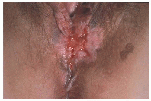
FIGURE 49.9 Invasive squamous cell carcinoma involving the perineum and perianal skin. Note the pigmented lesions of carcinoma in situ adjacent to the ulcerated invasive cancer.


Regional Lymph Nodes

It should be kept in mind that certain vulvar cancers, by virtue of their anatomic extent, may have access to lymphatics that bypass the groins. These include tumors that extensively involve the anus (particularly the anal canal or its surrounding tissue), the rectovaginal septum, the vagina above the lower third, and the proximal urethra. However, for the vast majority of vulvar cancers, the primary metastatic pathway is via the inguinal lymph nodes.


Sentinel Lymph Node Dissection

A large number of reports have been published over the past 25 years on sentinel lymph node detection in various cancers. The concept relies on the presumption that the sentinel lymph node is the initial site of metastatic disease and that lack of metastatic involvement of the sentinel lymph node reflects an absence of metastasis in the rest of the lymph nodes in the basin. Lymphatic mapping is the standard of care in the United States for the surgical treatment of patients with clinically early-stage melanoma and breast cancer.

In the case of invasive squamous cell carcinoma of the vulva, interest in sentinel lymph node was driven by the high incidence and significant morbidity of complications following full inguinal-femoral lymphadenectomy. Though the incidence of groin wound complications has been reduced by separate incisions, it still may occur in up to 30% of patients, and the risk is increased by patient comorbidities and adjuvant chemotherapy or radiation. Therefore, the use of a lesser surgical procedure such as sentinel lymph node dissection is particularly relevant to vulvar cancer.

The concept of sentinel lymph node dissection applies well to vulvar cancer as the tumor location is easily accessible to injection of both radiocolloid and blue dye, and the lymphatic drainage is predictable for most tumors. With lymphatic mapping, the pathologist has only a few lymph nodes to examine, allowing a more detailed examination. Techniques such as serial sectioning, immunohistochemical staining, and reverse transcriptase-polymerase chain reaction analysis can be applied, increasing the sensitivity of the examination and allowing the detection of micrometastases.

When using the combined technique for vulvar cancer patients, lymphatic mapping is nearly always successful in identifying sentinel lymph nodes (Fig. 49.10). Further, the false-negative rate (based on standard pathology) is very low. A meta-analysis by Hassanzade and colleagues showed that pooled sensitivity of 27 studies was 92% and a negative predictive value of 97%. A phase III trial by the GOG including tumors from 2 to 6 cm showed a sensitivity of 92.1% and a negative predictive value of 97.4% when a combination of blue dye and radiocolloid was used. Another multiinstitutional observational study by van der Zee et al. revealed a sensitivity of 94.1% and negative predictive value of 97.1%. A full inguinal-femoral lymph node dissection is reserved for those patients in which the sentinel node is positive or has not been successfully identified.

Sentinel lymph node mapping is a promising method to substantially reduce the morbidity of inguinal lymphadenectomy. It is widely used in the management of vulvar cancer today. However, many unanswered questions remain, including who the best candidates are for the technique, the role of frozen section, the role of immunohistochemistry, the role of lymphoscintigraphy, the reliability of an isolated lymphoscintigraphy-directed unilateral negative sentinel node with a midline lesion, the role of ultrasound, management of a patient with a microscopically positive sentinel lymph node, the incidence of “skip” metastases, the reliability of lymphatic mapping after prior excisional biopsy, and the optimal lymphatic mapping methodology for vulvar cancer patients.


Modifications on the Extent of Inguinal-Femoral Lymphadenectomy

Various modifications on the extent of lymphadenectomy have been examined. Before the introduction of sentinel lymph node biopsies, a technique involving limited resection of the inguinal lymph nodes in the management of superficially invasive vulvar cancer was reported by DiSaia and coworkers in 1979. The dissection they described is aimed at removal of the superficial lymph nodes above the cribriform fascia, mainly associated with the great saphenous and superficial epigastric veins. These lymph nodes were sent for frozen section analysis. If results were positive, a complete bilateral inguinofemoral lymphadenectomy was performed. In the 1979 study, DiSaia and colleagues also reported 79 cases of invasive squamous cell carcinoma of the vulva treated with radical vulvectomy and bilateral inguinal-femoral lymphadenectomy. In these cases, it was noted that the deep femoral lymph nodes were never positive in the absence of positive superficial inguinal lymph nodes. The purpose of a superficial dissection only is to reduce the morbidity of the groin lymphadenectomy. The dissection is less radical and resulted in only one groin breakdown in the 18 patients in the series of DiSaia and associates. The series was updated in 1989, and they reported no groin recurrences in 50 patients, 42 of whom had been followed for a median of 36 months.

A GOG study specifically studied the issue of superficial inguinal lymphadenectomy in patients with early carcinoma of the vulva. The study group included clinical stage I patients with tumor invasion of 5 mm or less and no capillary or lymphatic space involvement. A modified radical vulvectomy
and ipsilateral superficial inguinal lymphadenectomy were performed, and 121 patients were evaluable. These were compared with a historical control group in the GOG registry who had undergone radical vulvectomy with bilateral inguinofemoral lymphadenectomy. Nine patients in this study, or 7.3%, experienced groin recurrences versus no recurrence in the control group. Six of the groin recurrences were in the ipsilateral groin, and 5 of the 9 patients died of the recurrent vulvar cancer. The interpretation from this study was that superficial inguinal lymphadenectomy may not be adequate treatment even for early vulvar carcinoma. However, in a number of patients in this study, the tumors approached the midline; there is evidence that more medial tumors may have direct drainage to the deep inguinal lymph nodes. Another area of concern in this study is the high percentage of poorly differentiated tumors—almost twice as many as in the control group. Six of the nine groin recurrences in this study were from the poorly differentiated tumors. Whether poorly differentiated tumors are more likely to metastasize to deep inguinal or contralateral inguinal lymph nodes deserves further study. Subsequent additional retrospective data from large cancer centers also report a 5% to 10% incidence of groin relapse in patients with negative nodes from superficial inguinal lymphadenectomy.


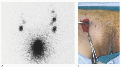
FIGURE 49.10 A: Preoperative lymphoscintigram 60 minutes after intralesional injection of technetium-99. B: Intraoperative blue node blue sentinel node in the right groin after injection of isosulfan blue dye into the primary vulvar cancer.

The technique of superficial lymphadenectomy of the inguinal nodes above the cribriform fascia has been largely replaced by sentinel lymph node biopsy, but the occurrences of groin recurrences after apparently negative superficial nodes in these studies remains concerning.

The benefit of sparing versus sacrificing the saphenous vein remains unclear. Various studies have found conflicting results on the reduction of the incidence of lymphedema. A randomized trial by the GOG showed that application of fibrin sealant at time of lymphadenectomy also did not reduce the incidence of lymphedema.


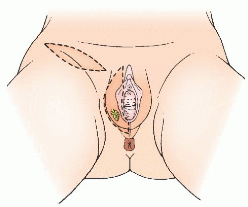
FIGURE 49.11 Unilateral lymphadenectomy in a patient with a focal right-sided small vulvar primary.


Unilateral Groin Lymphadenectomy

Unilateral lymphadenectomy only on the side of a unilateral vulvar tumor has been another modification of surgical treatment that has been used for selected patients (Fig. 49.11). In 1981, Iversen reported on 53 women with unilateral tumors and lymph node metastases. Eighty-three percent of these patients had only one positive ipsilateral node, 15% had bilateral positive nodes, and one patient had contralateral positive lymph nodes only. Other retrospective studies have confirmed these results. It has been the opinion of a number of authors that capillary or lymphatic space involvement by tumor may increase the
risk of contralateral nodal metastases. Patients with tumors approaching the midline or involving more medial structures (perineum, clitoral hood or clitoris, vagina, and labia minora) are at increased risk for contralateral lymph node metastases. The issue of unilateral groin lymphadenectomy was studied to some extent in 1992 by Stehman and associates in a GOG study. Patients with early disease and negative ipsilateral superficial inguinal lymph nodes were treated with ipsilateral superficial inguinal lymphadenectomy and a modified radical vulvectomy. A few patients in this study did have a bilateral inguinal lymphadenectomy because of midline involvement. A total of 121 patients were in the study, and 3 experienced contralateral inguinal lymph node recurrences. The vulvar lesions of these 3 patients ranged from 0.6 to 2.5 mm in depth of invasion, and all were poorly differentiated. Although lesion location was not given for these 3 cases, a large percentage of patients included in this study had lesions approaching the midline, as defined by involvement of the labia minora. Tumors with capillary or lymphatic space involvement were excluded from this study.

At present, unilateral inguinal-femoral lymphadenectomy appears to be a reasonable approach in a patient with a welllateralized early tumor that is well differentiated, with no capillary or lymphatic space involvement and with negative ipsilateral inguinal lymph nodes.


Pelvic Lymphadenectomy

During the late 1970s and early 1980s, several studies were published showing that carcinoma of the vulva metastasizes to the inguinofemoral lymph nodes before spreading to the pelvic lymph nodes. Extension of the groin lymphadenectomy to include removal of the pelvic lymph nodes continued to be performed in selected patients with positive inguinofemoral lymph nodes. A number of studies also showed that the 5-year survival rate of vulvar cancer patients with positive pelvic lymph nodes is less than 20%. A 1986 study by the GOG directed by Homesley compared pelvic lymph node dissection with groin and pelvic radiotherapy in patients with positive inguinofemoral lymph nodes. The study included 114 patients and showed no difference in morbidity between the two treatment arms and a better 2-year survival rate in the radiotherapy group (68% vs. 54%). The improved survival was seen in those patients with suspicious or grossly positive lymph nodes or those with more than one positive groin lymph node. There was no evidence that groin radiation therapy was beneficial to those patients with occult metastases and only one positive groin node. Review of the pattern of recurrence in that study suggested that adjuvant radiation was more effective largely because groin recurrences were reduced. The value of removing positive pelvic lymph nodes before radiotherapy is unknown. Pelvic lymphadenectomy is now rarely indicated in the treatment of vulvar cancer.


Omitting Groin Dissection for Superficial Disease

Extensive data support the contention that a subset of early vulvar carcinomas (carefully studied pathologically) can be identified that have an extremely low risk of nodal involvement. In a GOG study reported by Sedlis and associates, a subgroup (63 of 272) of patients with early disease was identified as having a zero incidence of lymph node metastases. This subset included nonmidline tumors with no capillary or lymphatic space involvement that were well differentiated or were grade 2 and limited to 2 mm in thickness. Other factors that have been studied for the purpose of identifying a low-risk group include tumor volume (which does not appear to have received further attention since Wilkinson’s report in 1985), tumors that are largely carcinoma in situ with very early stromal invasion and with a pushing rather than an infiltrative pattern, tumor diameter, squamous cell type, the presence of an inflammatory response, and tumor ploidy.

There seems to be general agreement that the patients for whom it would be most reasonable to omit the lymphadenectomy are those with tumor invasion less than or equal to 1 mm and less than 2 cm in diameter. However, certain tumors with less than 1 mm of invasion should still be considered for lymphadenectomy. As reported in the literature, three of nine patients with nodal disease or nodal recurrence in association with tumor invasion less than or equal to 1 mm had poorly differentiated cancers (Table 49.2). Thus, women with suspicious lymph nodes, poorly differentiated tumors, tumors with capillary or lymphatic space involvement, and perhaps those with multiple foci or broad areas of invasion or aneuploidy should still undergo lymphadenectomy. It is important to remember that even with superficial invasion, metastases to the nodes do occur. In the report by Sedlis and colleagues of 272 patients with invasion of 5 mm or less, the groin nodes were positive in approximately 20%.


Operative Technique


Radical Vulvectomy

With the classic en bloc resection, the inguinal specimens are mobilized toward the vulva as just described (Fig. 49.12). This operation is rarely employed now due to its morbidity and data on favorable oncologic outcomes with less radical surgery. From the point of completion of the inferior inguinal dissections, incisions are continued down along the labiocrural folds on each side and across the perineum, where they meet. A medial mucosal incision is made along the introitus extending through the anterior vestibule and around the urethral meatus.


FIGURE 49.12 Classic en bloc resection of the vulva and inguinal-femoral nodes through one interconnected incision.

When radical vulvectomy is performed through a separate vulvar incision, the same labiocrural, posterior, and mucosal
incisions are used, and the same vulvar tissue is excised (Fig. 49.13A). The superior incision extends from the top of the labiocrural folds as an inverted V, with the point above the base of the clitoris. As previously discussed, a variable amount of superior tissue (i.e., mons pubis) is removed, depending on the location and size of the lesion.


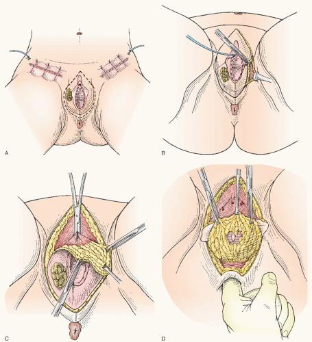
FIGURE 49.13 Radical vulvectomy with bilateral inguinal lymphadenectomy done through three separate incisions. A: Bilateral inguinofemoral lymphadenectomy through separate incisions is completed. Separate incision for radical vulvectomy is marked. B: Labiocrural incisions are extended to deep fascia of the urogenital diaphragm. C: Dissection proceeds dorsally off of the pubic bone. The vascular base of the clitoris is clamped, followed by transection and ligation. D: Perineal body and posterior vulvar tissues are dissected away from the anus.

The radical vulvectomy incisions may be modified somewhat depending on the location and extent of the tumor and the condition of the remaining uninvolved vulvar skin. The surgeon should attempt to attain at least a 2-cm margin of normal-appearing skin or mucosa around the tumor. To accomplish this, it may be necessary to excise a portion of the vagina,
anus, or distal urethra. For an anterior lesion, it is reasonable to spare the perineal body; but for a posterior lesion, it is important to incorporate radical resection of this area. For a lesion (especially superficial) in proximity to the urethral meatus or anus, it is reasonable to limit the margin of resection to 1 cm (but not less) to preserve these structures and their function.


FIGURE 49.13 (Continued) E: The vascular vestibular tissue along the sides of the vaginal tube is clamped. Transection and suture ligation to follow. F: Radical vulvectomy resection is completed. G: Closure of the vulvar wound is completed.

The labiocrural incisions are extended to the lateral margins of the deep fascia of the urogenital diaphragm (Fig. 49.13B). The internal pudendal vessels are ligated as they are encountered entering the vulva at about the 4 o’clock and 8 o’clock positions. Superiorly, the specimen is dissected off the pubic periosteum and adductor fascia. The vascular base of the clitoris is clamped and transected, and a transfixion suture ligature is placed (Fig. 49.13C). If deemed necessary, the attachment of the ischiocavernosus muscles can also be transected at this level. Dissection of the superior portion of the vulva off of the pubic bone and adductor fascia is completed and joined, in the midline, to the transvestibular mucosal incision above the urethra. Inferiorly, a variable portion of the perineum (and in some cases the anus) is dissected upward and cephalad toward the vaginal incision (Fig. 49.13D). Care is taken as the vaginal incision is approached above the perineum to avoid injury to the anal canal. Using the index finger or a large Kelly clamp, the surgeon separates the vaginal tube bluntly from the underlying soft tissue cephalad to the vestibular structures. The mucosal incision is then completed, separating the vagina (with the urethra) from the specimen. This dissection can be facilitated by splitting the specimen in the midline anteriorly or posteriorly. The remainder of the dissection off the
underlying deep fascia of the urogenital diaphragm is completed. Clamps and transfixion suture ligatures of no. 0 or 2-0 polyglactin are used during transection of the tissue along the side of the vaginal tube in the region of the vestibular bulbs (Fig. 49.13E).

After removal of the specimen, the wounds are copiously irrigated with antibiotic solution, and hemostasis is secured (Fig. 49.13F). After en bloc resection, the superior abdominal wall flap and the groin and vulvar thigh flaps are further mobilized as necessary to achieve closure of the wounds without tension. Any ischemic-appearing skin is excised. It is very useful, especially if there is an element of vaginal relaxation, to mobilize the lower vagina to facilitate wound closure. Any pelvic relaxation defects (i.e., cystocele, rectocele, loss of the posterior urethrovesical angle) are repaired at this time. To the extent that it is necessary or possible, the perineal body is also reinforced or reconstructed at this time.

The vulvar wound and the perineal area are closed with vertical mattress 2-0 delayed absorbable sutures (Fig. 49.13G). Depending on the amount of tissue that has been removed, the superior portion of an en bloc wound may be difficult to close. When done through a separate incision, closure of the vulvar wound is under much less tension, again because of preservation of the inguinal skin bridges. Careful attention must be paid to closure of the periurethral area. The urethra should be secured on a straight course without tension. A hood of skin above the urethra is also avoided because this can obstruct the path of the urinary stream and cause spraying.


Modified Radical Vulvectomy

As previously discussed, it is reasonable in most patients to manage the vulvar lesion locally with a modified radical vulvectomy. This operation consists of radical removal of the portion of the vulva containing the tumor and any skin, which is involved with high-grade VIN. It is performed with the techniques described earlier except that the excision is basically limited to removal of that particular part of the vulva. The lateral and deep tumor margins are not compromised by this operation. After the surgeon carefully demarcates a 2-cm radius of normal skin or mucosa around the tumor, an encompassing incision is designed that will readily close and be as cosmetically acceptable as possible (Fig. 49.14).


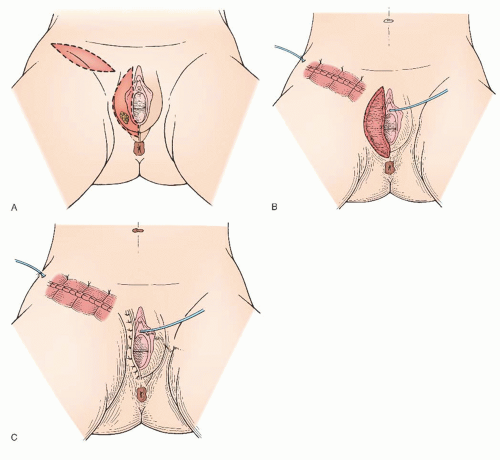
FIGURE 49.14 Modified right radical vulvectomy and unilateral right inguinal-femoral lymphadenectomy.


Lymphadenectomy


Sentinel Lymph Node Dissection

Sentinel lymph node detection is currently accomplished using two methods of lymphatic mapping: blue dye and radiocolloid. One to two milliliters of isosulfan blue dye (Lymphazurin) is injected superficially around the periphery of the primary tumor, and the blue-dyed lymphatic channels are followed. An incision is made over the anticipated location, and the dyed lymph node or nodes that have been stained by accumulating the blue dye are removed. Alternatively, the periphery of the tumor is injected with 400 mCi of technetium-labeled sulfur colloid 2 to 4 hours before surgery. A preoperative technetium scan (lymphoscintigraphy) is done, which may be helpful in confirming lymph node uptake, localization of target lymph nodes, unilateral versus bilateral lymphatic drainage, and identification of the unusual case of predominant drainage to pelvic lymph nodes. An intraoperative gamma counter is used to identify one or more sentinel lymph nodes (Fig. 49.15). The removed lymph nodes are checked with the gamma counter, and complete removal is assured when the radioactivity in the inguinal area returns to background levels. In practice, the two techniques are complementary and are frequently used together. The removed nodes are sent for frozen section, and the small skin incision in the groin is closed with absorbable sutures. Drains are not required. If the frozen section is negative, no further lymphadenectomy is required. However, if metastatic disease is detected, a complete inguinal-femoral lymphadenectomy is indicated.


Inguinal-Femoral Lymphadenectomy

An incision is made starting about 2 to 4 cm medial and about 2 cm caudal to the anterior superior iliac spine. The incision gradually curves downward just above the superior border of the inguinal ligament medially to the inguinal ring or about 2 cm below and 2 cm medial to the pubic tubercle. Unless there is a large clitoral lesion or palpably suspicious nodes, the mons pubis is spared, and separate incisions with a skin bridge are made as illustrated in Figure 49.16. For anterior lesions, such as those involving the clitoris, a portion of the mons pubis is included in the resection and the lymphadenectomy on one or both sides may be done en bloc with the radical vulvectomy as illustrated in Figure 49.12. From the lateral points, caudal incisions are carried medially so as to excise a strip of skin (optional) 2 to 4 cm in width. Excising an ellipse of the skin may reduce the likelihood of skin necrosis and facilitate more complete dissection of the groin. This incision is designed to extend from just below the fossa ovalis (this can generally be identified clinically as the area just medial to the femoral pulsation in the groin) to the top of the labiocrural fold above a point just medial to the external inguinal ring. In the presence of grossly positive inguinal lymph nodes, a wider resection of both the groin skin and fat is necessary to help ensure adequate tumor clearance. The separate groin skin incision may also be done vertically (with the leg), centered across the fossa ovalis, and about halfway between the femoral artery and pubic tubercle.


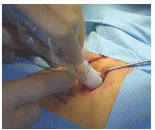
FIGURE 49.15 Intraoperative use of the gamma probe to identify the radioactive sentinel node.


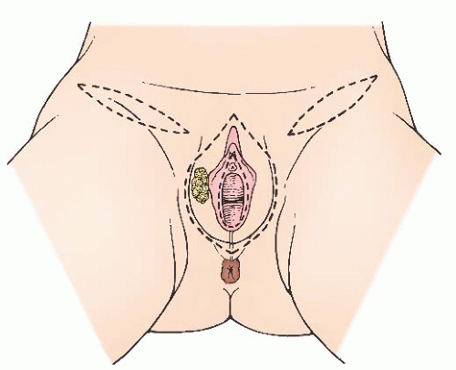
FIGURE 49.16 Separate incisions for bilateral lymphadenectomy done with a radical vulvectomy.

Leaving a layer of subcutaneous tissue with the skin, the superior incision is undermined so that the lymph node-bearing adipose tissue above the inguinal ligament and around the superficial circumflex iliac, as well as the superficial epigastric vessels, are included with the resection. These vessels are ligated or electrocoagulated as they are encountered. The superior dissection over the groin area is carried down to the superior border of the inguinal ligament (Fig. 49.17A and B). The midline aspect of the superior flap can be mobilized off the pubic bone and rectus fascia at this point to facilitate later closure of the wound without tension. Dissection of the block of inguinal tissue is carried inferiorly off of the inguinal ligament. The lateral corner is dissected medially off of the sartorius fascia. The inferior flap of the lower incision is also mobilized, especially medially, and the saphenous vein is identified as it enters the region of the femoral triangle (Fig. 49.17C). Accessory saphenous veins can also be seen entering this area. If it is to be sacrificed, the long saphenous vein is isolated and ligated with a free-tie and a transfixion ligature of 2-0 polyglactin (Vicryl). The vein with its surrounding block of lymph node-bearing tissue is dissected superiorly off of the sartorius and adductor fascia. Dissection of the block of inguinal tissue is continued from the three sides toward the fossa ovalis. As this area is approached, the overlying cribriform fascia is recognized, and the femoral artery pulsation can be palpated in the lateral aspect. En bloc dissection continues and includes the contents of the fossa ovalis (Fig. 49.17D). The area under the fascia lateral to the femoral artery should be left undisturbed as the femoral nerve lies lateral. There are no lymph nodes of consequence here, and avoidance of this area prevents injury to the femoral nerve and possibly reduces subsequent
lymphedema. Rather, resection of the cribriform fascia begins over the area of femoral pulsation, exposing the underlying femoral vessels. A few small branches of the femoral nerve are sacrificed during this dissection. The sheath of the femoral artery is incised along its anteromedial aspect from somewhere between the base of the fossa ovalis and the apex of the femoral triangle to its emergence from under the inguinal ligament. Branches, such as the external pudendal artery, are ligated as they are encountered. There is no purpose in dissecting under the artery or between the femoral artery and vein. Rather, the dissection that has been performed over the top of the artery is continued over the top of the vein, mobilizing the specimen to the medial aspect of the femoral vein. During this process, the saphenofemoral venous junction is identified, ligated with a 2-0 silk free-tie followed by a suture ligature for security, and transected, thus removing several centimeters of the saphenous vein with the specimen (Fig. 49.17E).


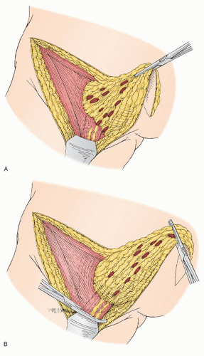
FIGURE 49.17 A: Inguinal-femoral lymphadenectomy. The corner of the groin specimen is dissected up from lateral to medially off the sartorius muscle. The lateral portion of fossa ovalis and cribriform fascia are exposed. B: The block of lymph node-bearing tissue has been mobilized, and the fatty lymphatic tissue overlying the saphenous vein is clamped.

An alternative method, as long as there are no adherent suspicious lymph nodes, is to dissect the saphenous vein free of the specimen so that it can be preserved, potentially reducing the risk of subsequent lymphedema. Complete dissection of the space medial to the vein is important because this is where most of the femoral groin nodes are located. The specimen is freed from the femoral vein medially, from the inguinal
ligament superiorly, and from the underlying pectineal fascia. Dissection is continued toward and off of the adductor longus fascia until the labiocrural fold is reached (Fig. 49.17F).


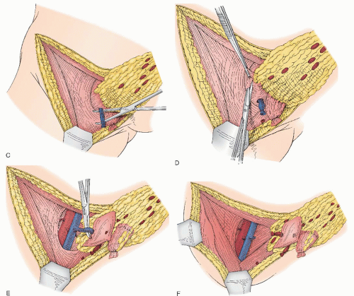
FIGURE 49.17 (Continued) C: The saphenous vein is identified, isolated, clamped, and divided near its entrance into the femoral triangle. D: Dissection of the femoral lymph nodes beginning with the separation of the cribriform fascia and the contents of the fossa ovalis from the anterior aspect of the femoral artery. E: The groin specimen containing the fatty nodal tissue above the cribriform fascia and the femoral nodes below the cribriform fascia are reflected medially exposing the femoral vessels and the insertion of the saphenous vein. F: The saphenous vein has been divided and ligated. The nodal tissue bundle is then completely excised and removed. The femoral triangle has been cleared of all nodal tissue.

Only gold members can continue reading. Log In or Register to continue

Stay updated, free articles. Join our Telegram channel

Jun 4, 2016 | Posted by in GYNECOLOGY | Comments Off on Malignancies of the Vulva

Full access? Get Clinical Tree

Get Clinical Tree app for offline access