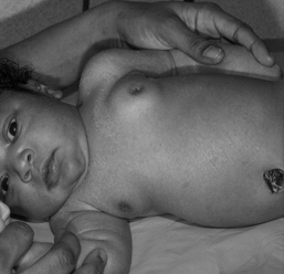
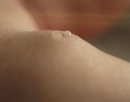
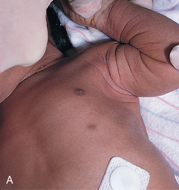
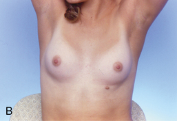
CHAPTER 16 Male and female breast development is similar until puberty; therefore, the examination of both boys and girls will be reviewed in this chapter along with pubertal changes expected for both genders. The primary intention of the breast exam in children and adolescents is to recognize normal variants, monitor development, and to identify nonmalignant pathology. Making assessment of the breast part of the routine physical examination in preadolescents gives the parent or caregiver and child an opportunity to voice any concerns and fosters a dialogue about reproductive health. It is important to respect privacy in performing the examination of the breast in all children and adolescents. Current evidence and recommendations related to the efficacy of breast examination focus on the detection of breast cancer, which is an exceedingly rare diagnosis in adolescents; therefore, child and adolescent breast exam recommendations are based on expert practice rather than evidence.1 A mammary ridge forms from the ectodermal layer on day 20 of embryonic life and extends from the forelimb to the hind limb. In the sixth week of fetal life, the nipple and areola form over a bud of breast tissue that is composed of the primary mammary ducts and a loose fibrous tissue or stroma. Fifteen to twenty-five secondary buds then develop and bifurcate into tubules, forming the basis of the duct system.2 Each duct, as it develops, opens separately into the nipple. At birth, the breasts of both male and female infants may be swollen because of the maternal estrogen effect (Figure 16-1). An unusual but normal finding in the newborn is the secretion of a milklike substance, also known as “witch’s milk,” for 1 to 2 weeks3 (Figure 16-2). Male and female breasts are similar until puberty, consisting of a small amount of breast tissue. Occasionally, a prepubertal male or female develops an enlargement of one or both breasts, which involves a soft, mobile, subareolar nodule of uniform consistency.2 Generally, the nipple and areola are not developed or pigmented with such an enlargement, and no associated signs of puberty are present. This condition usually regresses spontaneously within weeks to months, and in the absence of other secondary sexual characteristics, a biopsy is not indicated.2 If other signs of puberty appear, then these changes could be the first sign of precocious puberty, in which case referral and further diagnostic workup are indicated. FIGURE 16-1 Estrogen effect in the newborn. (From Shah BR, Laude TA: Atlas of pediatric clinical diagnosis, Philadelphia, 2000, Saunders.) FIGURE 16-2 Witch’s milk. (From Clark DA: Atlas of neonatology, ed 7, Philadelphia, 2000, Saunders.) Supernumerary nipples may arise from the mammary ridge and be present at birth (Figure 16-3, A). They are often raised, generally require no treatment, and become imperceptible over time. There is a weak association between supernumerary nipples and renal and cardiovascular abnormalities in white newborns.3 In females, supernumerary nipples will on rare occasion develop a small amount of breast tissue during puberty (see polymastia in Table 16-2; see Figure 16-3, B). The adolescent may elect to have cosmetic surgery for removal of the supernumerary breast tissue. Widespread nipples are defined as a nipple spread of greater than 25% of the chest circumference and may be associated with congenital disorders such as Turner syndrome.3 FIGURE 16-3 A, Supernumerary nipple. B, Supernumerary nipple located in the inframammary fold of the left breast of an adolescent female. (A from Eichenfield L, Frieden IJ, Zaenglein AL: Neonatal dermatology, ed 2, Philadelphia, 2009, Saunders; B from van Aalst JA, Sadove M: Treatment of pediatric breast problems, Clin Plast Surg 32(1):65-78, 2005.) At puberty, the ductal and periductal mesenchymal breast tissue of boys proliferates under the influence of estrogens, with later involution as testicular androgens rise to adult levels.2 Male estradiol levels triple during puberty, and androgens ultimately increase 30 times. If peak estrogen levels occur before androgen levels, the result is gynecomastia, a benign increase in glandular and stromal breast tissue in pubertal males (Figure 16-4). Most circulating estrogens are produced outside of the testes, and an increase in fatty tissue, as in obesity, may lead to an increase in estrogen levels and a higher incidence of gynecomastia (Box 16-1). At 14 years of age, 64% of adolescent males have some degree of gynecomastia, with only 4% of adolescent males having severe gynecomastia that persists into adulthood.2 Approximately 50% of males experience the onset of gynecomastia at Tanner stage 2 of male genital development, another 20% at Tanner stage 1, 20% at Tanner stage 3 of male genital development, and 10% beginning at Tanner stage 4 (see Chapter 15). In general, adolescent males can be reassured that most cases of physiological gynecomastia will resolve spontaneously within 6 months, although it can persist up to 2 years after attaining sexual maturity.4 Treatment alternatives for persistent gynecomastia, including pharmacological and surgical options, may be appropriate to discuss with youth and families when this condition does not resolve or is particularly distressing. Gynecomastia, although a normal variant, is also present in relation to hormone imbalances due to thyrotoxicosis, cirrhosis, adrenal and testicular neoplasm, primary hypogonadism, chromosome abnormalities such as Klinefelter syndrome, and severe malnutrition.2 In addition, prescription drugs such as cimetidine, ketoconazole, metronidazole, isoniazid, digoxin, spironolactone, phenothiazines, and some illicit drugs (e.g., marijuana, anabolic steroids, amphetamines, and opiates) can cause gynecomastia.4 Thelarche, or the beginning of female breast development, is usually the first sign of puberty in girls and occurs between 8 and 13 years of age, on average at the age of 11.2 years1 (Table 16-1 and Figure 16-5). Full breast development at Tanner stage 5 signals the end of puberty in females. Breast development during puberty involves both multiple hormones and the binding of hormones to breast tissue. Estrogen, especially estradiol, influences ductal development, whereas progesterone influences additional lobular alveolar development.2 Thyroxine and corticosteroids are also involved. TABLE 16-1 Female Breast Development Sexual Maturity Rating Data from Neinstein LS: Adolescent health care: a practical guide, ed 5, Philadelphia, 2008, Lippincott Williams & Wilkins.
Male and female breast
Embryological development
Physiological variations
Physiological variations in the male breast

Physiological variations in the female breast
Tanner Stage/Sexual Maturity Rating (SMR)
Breast Findings
Areola and Nipple Findings
Tanner stage 1
Prepubertal; no glandular tissue
Conforms to general chest line
Tanner stage 2
Breast bud; small amount of glandular tissue
Areola widens
Tanner stage 3
Larger and more elevation; extends beyond areolar parameter
Areola continues to enlarge but remains in contour with breast
Tanner stage 4
Larger and more elevation
Areola and papilla form a mound projecting from breast contour (half of teens; in some cases persists into adulthood)
Tanner stage 5
Adult size (variable)
Areola part of breast contour, nipple projecting above areola
![]()
Stay updated, free articles. Join our Telegram channel

Full access? Get Clinical Tree


Male and female breast
Only gold members can continue reading. Log In or Register to continue
