Fig. 5.1
a Plain X-ray showing a large soft tissue density in the axillary region in a patient with large cystic hygroma. b Clinical photograph of the same patient in showing a large axillary cystic hygroma extending downwards

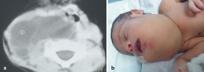
Fig. 5.2
a CT scan showing a large cervical lymphangioma. b clinical photograph showing a very large cervical lymphangioma

Fig. 5.3
Clinical photograph showing a large cervical lymphangioma in an older child
Lymphangiomas can however appear at any age.
This is specially so at sites other than the head and neck including the abdomen and mediastinum where they can attain a large size before being apparent clinically.
Sometimes, they can attain a large size as a result of infection or hemorrhage. This can cause pressure symptoms or life-threatening complications at sites like the mediastinum (Figs. 5.3, 5.4, 5.5, 5.6, and 5.7).

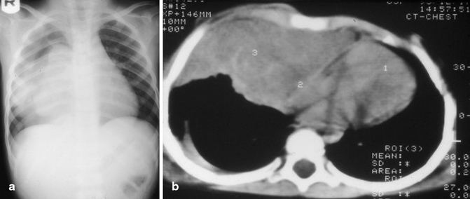
Fig. 5.4
a Chest ray showing a soft-tissue density in the right side of the chest and b CT scan of the chest for the same patient showing a large intrathoracic lymphangioma

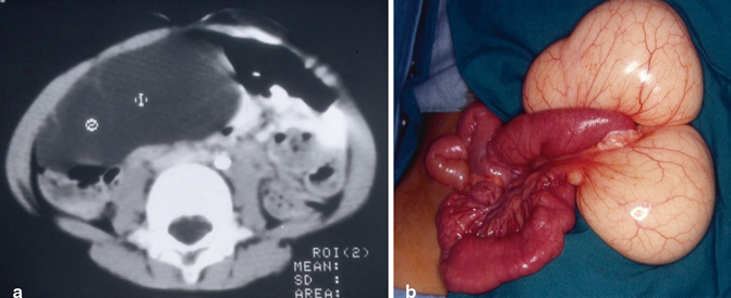
Fig. 5.5
a CT scan of the abdomen showing a large intra-abdominal lymphangioma and b intraoperative photograph showing a large mesenteric lymphangioma

Fig. 5.6
Intra-operative photograph showing a macrocystic lymphangioma arising from the abdominal wall

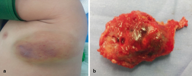
Fig. 5.7
a A clinical photograph showing lymphangioma of the chest wall. Note the spontaneous bleeding into the lymphangioma and b a clinical photograph of the resected lymphangioma. Note the bleeding inside the lymphangioma
An important observation is the sudden appearance of cervical lymphangioma as a result of bleeding. This causes diagnostic confusion and the final diagnosis is confirmed only intraoperatively and by histology.
The possibility of lymphangioma should always be considered in children with sudden appearance of a cervical swelling.
Classification
Lymphangiomas have traditionally been classified into three subtypes:
Capillary.
Cavernous lymphangiomas.
Cystic hygroma.
A fourth subtype the hemangiolymphangioma is also recognized.
This classification is based on their microscopic characteristics.
Capillary lymphangiomas are composed of small, capillary-sized lymphatic vessels and are characteristically located in the epidermis.
Cavernous lymphangiomas are composed of dilated lymphatic channels and characteristically invade surrounding tissues.
Stay updated, free articles. Join our Telegram channel

Full access? Get Clinical Tree


