11.2 Low birth weight, prematurity and jaundice in infancy
Principles of care
Care of the sick newborn is often complex, and requires specialized training and equipment. However, remembering the basic principles will allow you to provide emergency care for the sick newborn, regardless of diagnosis, until specialized help is available:
• Keep the baby pink. Initial resuscitation should follow the usual ABC guidelines (see Chapters 5.15.2 and 11.1). After that, many babies will maintain breathing with supplemental oxygen until more sophisticated respiratory support is available. If an oxygen saturation monitor is available, give only enough oxygen to keep a preterm baby’s oxygen saturations between 86% and 94%, and a term baby’s saturations above 95%.
• Keep the baby warm. Cooling increases the baby’s oxygen and glucose requirements and is associated with increased mortality. Dry the baby and put a hat on to reduce heat loss while you are assessing other problems. Use a radiant heater, electric blanket or incubator if available.
• Keep the baby fed. Sick babies are at risk of hypoglycaemia, which can cause brain damage. If milk feeds are not possible, give intravenous 10% dextrose 60 mL/kg daily (2.5 mL/kg hourly).
• Consider infection. Almost any signs and symptoms of illness in the newborn can be caused by infection, and untreated septicaemia can cause death within hours. If specialized care is likely to be delayed by more than an hour or two, take blood cultures if possible and give intravenous or intramuscular antibiotics.
Definitions
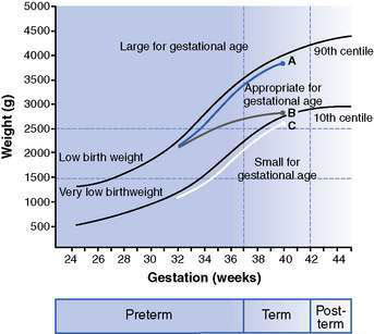
Babies are commonly classified into groups associated with different disease patterns and different outcomes (Fig. 11.2.1). These include:

Fig. 11.2.1 Common definitions of size at birth, illustrating the difference between intrauterine growth restriction (IUGR) and small for gestational age (SGA). Baby A is an appropriately grown term baby. Baby B is also born with an appropriate size for gestational age (AGA), but has suffered reduced intrauterine growth compared with baby A and thus has IUGR. Baby C has had normal intrauterine growth, but is born SGA.
The premature infant
Causes of preterm birth
Although there are many risk factors for preterm birth (Box 11.2.1), approximately half of preterm births occur in the absence of recognized risk factors. Survival rates increase and the incidence and severity of complications all decrease with increasing gestational age and birth weight (Figs 11.2.2, 11.2.3, Table 11.2.1).

Fig. 11.2.2 Survival of preterm infants admitted to neonatal units according to (A) birth weight and (B) gestational age.
(Source of data: Australian and New Zealand Neonatal Network (ANZNN) 2009 Report of the Australian and New Zealand Neonatal Network 2006. ANZNN, Sydney.)

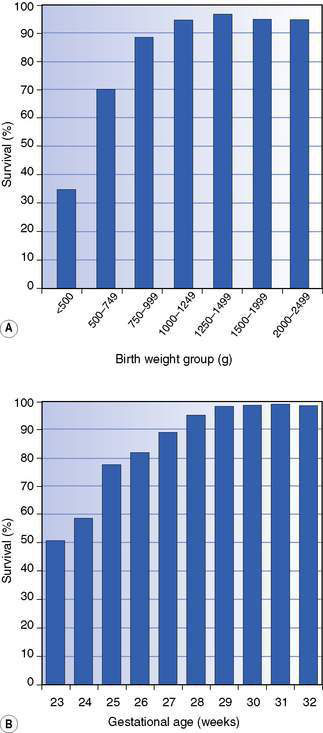
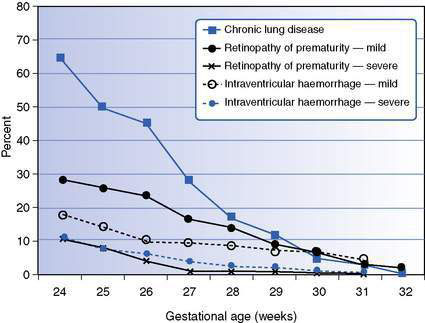
Fig. 11.2.3 Frequency of complications of prematurity in babies admitted to neonatal units according to gestational age.
(Source of data: Australian and New Zealand Neonatal Network (ANZNN) 2009 Report of the Australian and New Zealand Neonatal Network 2006. ANZNN, Sydney.)
Table 11.2.1 Complications of preterm birth
Common | Rare except in very low birth weight | |
---|---|---|
Early | ||
Respiratory | Respiratory distress syndrome | |
Apnoea | ||
Cardiac | Patent ductus arteriosus | |
Neurological | Periventricular haemorrhage | |
Periventricular leukomalacia | ||
Hepatic | Hypoglycaemia | Hyperglycaemia |
Hyperbilirubinaemia | ||
Renal | Hyponatraemia | Hyperkalaemia |
Metabolic acidosis | ||
Gastrointestinal | Feeding problems | Necrotizing enterocolitis |
Other | Anaemia | |
Infection | ||
Poor thermoregulation | ||
Late | Delayed growth | Retinopathy of prematurity |
Chronic lung disease | ||
Neurodevelopmental delay |
George is a 900-g (extremely low birth weight) baby born to a 16-year-old mother who received no prenatal care. His mother was admitted after the membranes ruptured and she began to have contractions. She did not remember the date of her last menstrual period and had not had any antenatal ultrasound scans. She smoked a pack of cigarettes per day during the pregnancy. At delivery, George had poor respiratory effort and marked retractions so he was intubated in the delivery room and brought to the neonatal intensive care unit. He required moderate ventilator settings and 50% oxygen. Chest X-ray showed a diffuse ground-glass appearance with air bronchograms consistent with respiratory distress syndrome. A dose of surfactant was given through the endotracheal tube. Gestational age was estimated at approximately 27 weeks based on the Ballard examination, which assesses physical and neuromuscular development. Based on this estimated gestation, the infant’s weight, length and head circumference were all at the 25th centile, and were appropriate for his gestational age.
Short-term complications of prematurity
Respiratory
Respiratory distress syndrome
Respiratory distress syndrome is also called surfactant deficiency syndrome. Immaturity of the respiratory system with surfactant deficiency results in respiratory distress. This is managed with oxygen, nasal continuous positive airway pressure (NCPAP) or, when more severe, surfactant administration and mechanical ventilation. Corticosteroids given to the mother before preterm birth reduce the incidence and severity of respiratory distress syndrome.
Periodic breathing, apnoea of prematurity
Premature babies commonly experience periodic breathing due to immaturity of the respiratory centres of the brain. Cessation of breathing persisting for more than 20 seconds, or less than 20 seconds if there is associated bradycardia or desaturation, is termed apnoea. Premature babies require cardiorespiratory and pulse oximetry monitoring to detect apnoea and associated bradycardia or desaturation. Apnoea of prematurity occurs in almost all extremely premature babies and usually improves around 34–36 weeks’ postmenstrual age. Pharmacological treatment includes methylxanthines such as caffeine or theophylline, which improve diaphragmatic contraction and stimulate the respiratory centres. NCPAP is also helpful, partly by reducing any obstructive component to the apnoea and reducing the work of breathing. If apnoea is severe, the baby may have to be ventilated mechanically. Apnoea can also be caused by many other complications of prematurity, such as infection, neurological problems, anaemia, hypoxia, patent ductus arteriosus and upper airway obstruction, so these need to be considered in babies experiencing apnoea.
Cardiac
Patent ductus arteriosus
Before birth, the ductus arteriosus diverts blood from the right ventricle away from the lungs to the aorta. After birth it normally closes functionally within a few days. In premature babies, closure may be delayed, leading to left-to-right shunting of blood from the aorta through the ductus to the lungs. This results in pulmonary congestion, worsening lung disease and decreased blood flow to the gastrointestinal tract and brain. These changes have been implicated in the pathogenesis of necrotizing enterocolitis and intraventricular haemorrhage. A significant patent ductus arteriosus (PDA) is often clinically silent, or there may be a continuous heart murmur, hyperdynamic precordium, bounding pulses and widened pulse pressure. Diagnosis is made by echocardiography. A significant PDA may be treated by giving prostaglandin inhibitors (indometacin or ibuprofen). If these are unsuccessful, surgical ligation may be necessary.
Neurological
Intraventricular haemorrhage
This is due to bleeding from the immature capillary bed of the germinal matrix lining the ventricles, often within the first 48 hours after birth. Risk factors include asphyxia and changes in cerebral blood flow due to hypotension or rapid intravenous fluid infusion. Intraventricular haemorrhage is diagnosed by cranial ultrasonography and varies in severity from grade I intraventricular haemorrhage (germinal matrix haemorrhage) to grade IV (intraparenchymal haemorrhage). Although lower grades have a good prognosis, grades III and IV intraventricular haemorrhage are often associated with later hydrocephalus and neurological abnormalities such as cerebral palsy.
Periventricular leukomalacia
This is an uncommon problem, characterized by ischaemic necrosis of the white matter surrounding the lateral ventricles. Periventricular leukomalacia is diagnosed on head ultrasonography, usually at 4–6 weeks of age. It often results in cerebral palsy.
George improved significantly after the administration of surfactant. He was extubated and was placed on nasal continuous positive airway pressure (NCPAP) by age 36 hours. Caffeine was given before extubation in anticipation of apnoea of prematurity. Small feedings of expressed breast milk were started on day 1. Electrolytes were monitored closely because of the potential of large insensible water losses.
On day 4, George began to deteriorate with increasing apnoeas and respiratory distress. A continuous murmur was noted along the left sternal border. The pulses were bounding and the precordium was very active. Echocardiography confirmed the presence of a large patent ductus arteriosus and a course of indometacin was commenced.
George did well during the next week. At 2 weeks of age, he again developed worsening apnoea with feed intolerance and temperature instability. A full blood count revealed anaemia and an increase in immature white cells. Blood cultures, a lumbar puncture, chest X-ray and bladder tap were performed. George was treated with a blood transfusion and antibiotics, with gradual improvement during the next 2 days.
Hepatic
Hypoglycaemia is common because of decreased glycogen stores and increased glucose requirements in premature babies.
Hyperglycaemia can also occur in VLBW babies because of high glucose infusion rates, reduced insulin secretion and impaired insulin sensitivity.
Hyperbilirubinaemia is common and due to hepatic immaturity coupled with a shorter half-life of red blood cells. Premature babies require treatment at lower bilirubin levels than term babies because their low albumin levels and immaturity of the blood–brain barrier place them at greater risk of bilirubin encephalopathy.
Renal
Immaturity of the kidneys results in a poor ability to concentrate or dilute the urine. This may be aggravated by immature skin leading to high insensible water losses, contributing to:

Stay updated, free articles. Join our Telegram channel

Full access? Get Clinical Tree

