Lid and Lacrimal System Disorders
Gregg T. Lueder
THE EYELIDS
The eyelids play an important role in maintaining ocular health and good vision. The mechanical action of the eyelids sweeping over the globe brings in fresh, lubricating tears and removes debris. The edge (margin) of the two upper eyelids and two lower eyelids should be symmetric. The upper eyelid margin should rest at the superior edge of the cornea or just over the superior iris, so as not to block any visual input. The eyelid margins normally rest against the globe, and the eyelashes are directed outward.
CONGENITAL ANOMALIES OF THE EYELID
Except for ptosis (drooping of the eyelids, blepharoptosis), which will be discussed below, congenital anomalies of the eyelids are rare. The most severe anomaly is cryptophthalmos, which presents in the newborn with complete fusion of the eyelids. The underlying eyeball is also usually malformed, and the visual prognosis is poor. Eyelid coloboma is a discrete area in which eyelid tissue is missing, most commonly on the upper eyelids, appearing as a notch or rectangular defect of the margin where there will be no lashes. Anomalies of the cartilage normally present inside the eyelid (tarsal plate) or eyelid muscles may result in congenital entropion (in-turning of the eyelid) or ectropion (out-turning of the eyelid). Congenital tarsal kink is a severe variant of ectropion, in which the upper eyelid appears to bend outward. Surgery for these conditions is indicated if they produce corneal irritation (due to eyelashes rubbing against the cornea in entropion) or exposure damage (due to inadequate corneal cover and lubrication in ectropion). A relatively minor eyelid abnormality is ankyloblepharon, which results from incomplete separation of the eyelids during embryologic development. Infants usually present with one or more fine strands of adherent tissue between the upper and lower eyelids, with secondary inability to open the eyes (eFig. 589.1  ). This can usually be treated successfully in the ophthalmologist’s office with simple cutting of the tissue.
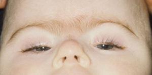
Epiblepharon is a relatively common congenital eyelid anomaly. It results from an extra fold of skin below the lower eyelid margin, which may cause the eyelids and lashes to rotate in toward the eye (eFig. 589.2  ). It is most commonly seen in Asian children. If the lashes rub against the cornea, affected infants may develop symptoms of ocular irritation, overflow tearing, and excess mucus formation. The symptoms of this disorder may be confused with those of nasolacrimal obstruction, and it is important to differentiate these two entities by inspecting the eyelid margin. Epiblepharon may spontaneously improve in the first 6 to 12 months of life, and conservative treatment with topical lubricants may provide symptomatic relief. If the condition does not improve, surgery to evert the eyelid margin is usually successful.1
PTOSIS
 CONGENITAL PTOSIS
Congenital ptosis usually results from under-development of the eyelid levator muscle. The condition may be bilateral or unilateral and may be isolated or associated with other ocular or systemic disorders. Unilateral ptosis is more likely to be isolated. Bilateral ptosis is more likely to be associated with other problems. Etiologies for bilateral ptosis include familial (usually autosomal dominant), chromosomal (eg, Turner syndrome), teratogenic (eg, fetal alcohol syndrome), and syndromic. Both bilateral and unilateral ptosis also frequently occur in otherwise normal children, in which case there is often no identifiable causative factor.
Ptosis presents with the upper eyelid resting in a lower position than normal on the eye. It may range from mild, with the eyelid 1 to 2 mm lower than normal, to severe, in which case the eyelid may cover the whole cornea. In congenital ptosis, the eyelid crease is usually absent, because the crease normally forms from fibers of the levator muscle (which is hypoplastic in these patients) attaching to the eyelid skin. The residual levator muscle in ptosis is also usually stiff, which may limit full closure of the lid.
In rare instances, children may have pseudoptosis. In this condition, the eye with the lower eyelid initially appears to be abnormal, but the asymmetry is actually due to proptosis (anterior displacement) of the contralateral eye, where the upper eyelid is somewhat retracted.
Approximately 5% of children with congenital ptosis have the Marcus-Gunn jaw-winking phenomenon. In this condition, the fibers that innervate the levator muscle are aberrantly connected to fibers that innervate the masseter muscle. This produces a synketic elevation of the eyelid during jaw movements. In infants, this is usually noted while they suck during feeding, and in older children, it can be seen by asking the child to move the jaw laterally. This usually manifests as a baseline ptosis with intermittent elevation or even retraction during jaw movements, but it may also manifest as a baseline normal eyelid position, with intermittent widening of the palpebral opening during jaw movement.
The primary visual problem associated with ptosis is amblyopia, which may occur for two reasons.2 First, if the eyelid margin rests at or below the pupil, it may interfere with vision. In unilateral cases, this may cause the child to favor the normal eye. Second, the mechanical weight of the eyelid may induce astigmatism (asymmetric curvature) of the cornea, creating a blur that also causes the child to favor the opposite eye. In mild cases (1 to 2 mm), ptosis usually does not produce any visual or appearance problems. In moderate cases (the ptotic lid is 3 to 5 mm below normal level), children will often adapt compensatory strategies to improve their vision. Once affected infants have adequate neck control, they often tilt their head back in order to look beneath the droopy eyelid (chin lift). In addition, children will often attempt to use the frontalis muscle to assist in lifting the eyelid, producing contraction of this muscle and, in unilateral cases, asymmetric elevation of the eyebrows (Fig. 589-1). In severe cases, in which the lid covers most of the cornea, there is a great risk of deprivation amblyopia, which may be irreversible if the ptosis is not corrected early.
Treatment of ptosis involves surgical elevation of the eyelids. Amblyopia treatment with patching and glasses for astigmatism is also often necessary. Severe ptosis needs to be corrected within the first few months of life due to the risk of amblyopia. Timing of surgery for moderate ptosis depends on the presence of amblyopia and compensatory head postures. If these are present, early surgery is indicated. If not, surgery is often delayed until age 4 to 5 years. Children with mild ptosis often do not require surgery.
 ACQUIRED PTOSIS
Compared to the incidence of congenital ptosis, acquired ptosis is relatively uncommon in children, and the differential diagnosis is large. Acquired ptosis rarely presents as an isolated phenomenon, and an etiology can often be identified by the history and associated findings. Possible etiologies include myogenic, neurogenic, inflammatory, infectious, and mass lesions.
Myasthenia gravis is a myogenic disorder that results from antibodies directed against acetylcholine receptors, which may initially present with ptosis.3 This can occur in the neonatal period and should be considered in the differential diagnosis of congenital ptosis, but it more commonly presents at an older age. Historical features that suggest this diagnosis include worsening of the symptoms with fatigue and intermittent diplopia (due to involvement of the extraocular muscles). Diagnostic tests include electromyography and serum anticholinesterase antibodies, and administration of edrophonium or neostigmine can improve symptoms.
Another myogenic disorder that may initially present with ptosis is chronic progressive external ophthalmoplegia (CPEO). This is usually due to mitochondrial dysfunction that affects the levator and extraocular muscles. Sporadic and inherited forms occur, and the ocular findings of CPEO may be seen in several diseases associated with mitochondrial dysfunction. One of these is Kearns-Sayre syndrome, which is associated with CPEO, pigmentary retinopathy, and cardiac conduction abnormalities. Children with CPEO should be evaluated by a cardiologist.
Neurogenic causes of ptosis include third cranial nerve palsy and Horner syndrome. The third cranial nerve innervates the eyelid levator muscle, four of the six extraocular muscles, and the iris muscles that constrict the pupil. In a complete third nerve palsy, the eyelid is ptotic, the eye is turned out and down (because the only residual functioning extraocular muscles are the lateral rectus muscle, which pulls the eye out, and the superior oblique muscle, which pulls the eye down), and the pupil is dilated (Fig. 589-2). In certain conditions, only the superior division of the third nerve is involved, in which case only innervation of the eyelid and superior rectus muscle are involved, producing ptosis and downward deviation of the eye.
Stay updated, free articles. Join our Telegram channel

Full access? Get Clinical Tree


