Fig. 6.1
Schematic representation of the room setup (P patient, AV audiovisual equipment, N scrub nurse, I instrument trolley S surgeon, A camera holder)
Anesthesia
Endotracheal intubation is required in all cases using either a cuffed or reinforced endotracheal tube, securely fastened. This is to prevent tube dislodgement when the child is positioned prone for the surgery. Perioperative and postoperative analgesia is provided by preemptive local infiltration of the planned incisions with 0.25 % bupivacaine.
Operation
Retroperitoneoscopic Nephrectomy (Video 6.1)
1.
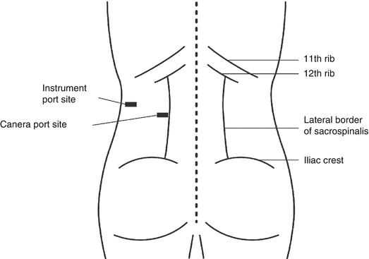
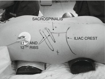
The patient is positioned fully prone under general anesthesia. Other approaches including the lateral and anterolateral approach are also popular. The exposed dorsal and lateral aspects of the trunk are prepared and draped in a sterile manner. Topographic landmarks and anticipated port sites are marked as shown (Figs. 6.2 and 6.3).


Fig. 6.2
Schematic representation of port position

Fig. 6.3
Preoperative demonstration of landmarks and port sites
2.
Creation of retroperitoneal space outside Gerota’s fascia by a technique described by Gill [8]. Several commercial balloons are available for creation of the retroperitoneal space. However, the author prefers a simple and inexpensive balloon made by securing the finger of a sterile surgical glove to the end of a 12 Fr Jacques catheter with a silk tie. The catheter is connected to a 3-way tap and a 50 ml luer-lock syringe. Depending upon the size of the patient, 100–250 ml of air is injected slowly to develop the retroperitoneal space. The system is left inflated for 2 min to promote hemostasis and then deflated and withdrawn.
3.
Insertion of primary and secondary ports: a 6 mm Hasson cannula is inserted into the port site, followed by insufflation of the retroperitoneum with CO2 to pressure of 10–12 mmHg. The Hasson port is secured by a suture to the skin. A 5 mm instrument port is placed under direct vision below the tip of the 11/12th rib and above the iliac crest. A second working port (5 mm) can be placed through the paravertebral muscles although in the author’s experience the nephrectomy can be performed using a single working port [9, 10].
4.
Exposure of the kidney: Gerota’s fascia is incised longitudinally adjacent to the posterior abdominal wall using scissors. The adventitious tissue is divided to gain adequate exposure and working space for the procedure.
5.
Exposure of the hilum: the kidney is dissected commencing at the apex and along the medial aspect. The lateral and inferior attachments are not divided at this stage as they anchor the kidney in position and aid in exposure of the hilar vessels.
6.
Division of the vascular pedicle: the vessels are divided between hemoclips or with a Ligasure® when the vessels are less than 8 mm in diameter. A minimum of three clips should be applied on all vessels, with at least two clips remaining behind on the proximal stump of the divided vessel.
7.
Ureteric division: the ureter is traced as far into the pelvis as is necessary. In cases of reflux-associated nephropathy, the ureter may be ligated with an endoloop or transected without ligation and the bladder drained with a urethral catheter for 48 h.
8.
The remaining attachments of the kidney are divided using a combination of blunt dissection, monopolar diathermy, and/or the Ligasure®. In the case of a large multicystic dysplastic kidney, complete intracorporeal mobilization can be technically difficult and time-consuming and risks creating a tear in the closely attached peritoneum. In such cases, after all vessels have been divided and the cysts decompressed, the kidney can be withdrawn via the camera port incision and the remainder of the dissection completed in an extracorporeal manner.
9.


Specimen retrieval: the specimen may be removed via the camera port depending upon the size. A multicystic dysplastic kidney or hydronephrotic kidney may be decompressed by aspiration and withdrawn directly via the camera port wound. A larger specimen may be retrieved after engaging it in a 10 mm Endopouch retrieval device and removing it piecemeal with sponge forceps.
Stay updated, free articles. Join our Telegram channel

Full access? Get Clinical Tree


