Fig. 16.1
Port placement for laparoscopic-assisted bladder augmentation using three ports
While there is no ideal bowel segment for augmentation cystoplasty, ileum is clearly the most common segment utilized due its compliance and minimal mucous production. The first step towards selecting an appropriate segment of ileum is identification of the appendix and ileocecal valve. Identifying these landmarks can be challenging in the presence of adhesions, commonly from a ventriculoperitoneal shunt in this population. If a shunt is present, then it is usually easy to move the shunt away from the site of dissection so as to minimize the risk of future infections.
The use of bowel graspers and bed positioning can help expose the appendix and cecum. Retracting the appendix anteriorly on stretch should expose the avascular line of Toldt and guide mobilization of the right colon. Electrocautery along the lateral edge of the right colon mobilizes the cecum and distal ileum in order to reach the pelvis. The appendix can be marked by leaving a grasper clamped to the distal tip.
While a Pfannenstiel incision allows for excellent exposure to the pelvis and bladder, identification of the appendix through this incision alone can be challenging. Prior laparoscopy eases identification and facilitates cephalad dissection for maximal mobility of the bowel into the pelvis. The distal 15–20 cm of ileum is spared in order to prevent steatorrhea and vitamin B12 deficiency. The remainder of the procedure should be conducted identically to a traditional open approach.
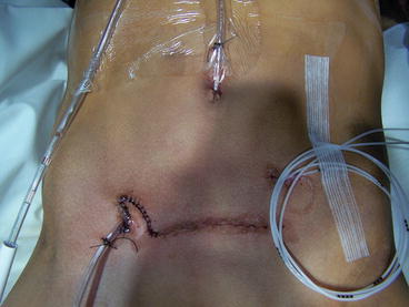
At the conclusion of the procedure, the right lower quadrant port site can be incorporated into the lateral edge of the Pfannenstiel incision, the umbilical incision is either buried within the umbilicus or used as a Mitrofanoff channel, and the 5 mm left lower quadrant incision may be used as the wound soaker exit. A suprapubic tube and wound soakers exit through the left lower quadrant (see Fig. 16.2).


Fig. 16.2
Final appearance of closure and tube placement
Postoperative Management/Acute Complications
All patients following enterocystoplasty are initially kept nil by mouth. The use of nasogastric decompression is generally not necessary and may introduce an unnecessary aspiration risk, particularly in this patient population [8, 10]. Postoperative electrolytes should be checked to ensure appropriate fluid management. Continuous bladder drainage should be maintained in addition to periodic irrigation twice daily to avoid occlusion by blood clot or mucous production. Clamping of catheters can be commenced after 10 days, followed by intermittent catheterization, usually after 4 weeks.
Acute complications following augmentation cystoplasty include bleeding, infection, urinary leak, and metabolic abnormalities. Bowel obstruction may occur in approximately 5 % of patients following augmentation [1]. Laparoscopy adds minimal morbidity to these known complications of open augmentation. Any enterotomy should be recognized and may be addressed either laparoscopically or open. Port-site hernia is a rare potential morbidity.
Results/Late Complications
Ileocystoplasty is a reliable method to increase bladder capacity and decrease intravesical storage pressures with a less than 10 % need for additional augmentation work [11]. Bowel will continue to display peristalsis or mass contraction, but this should be minimized by detubularization. The native bladder should be widely opened to avoid stenosis of the anastomosis.
Stay updated, free articles. Join our Telegram channel

Full access? Get Clinical Tree


