Fig. 11.1
Schematic representation of the room setup. P patient, AV audiovisual equipment, N scrub nurse, I instrument trolley, S surgeon, A camera holder

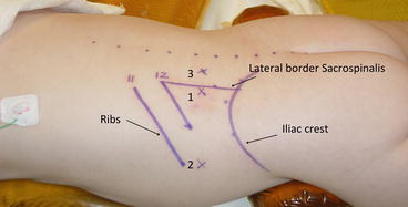
Fig. 11.2
Schematic representation of port position. 1 Camera port, 2 First instrument port, 3 Second instrument port
Anesthesia
Endotracheal intubation is required in all cases using either a cuffed or reinforced endotracheal tube that is securely fastened to prevent tube dislodgement when the child is positioned prone for the surgery. Preoperative and postoperative analgesia is provided by preemptive local infiltration of the planned incisions with 0.25 % bupivacaine.
Operative Technique
General Principles
The operative strategy is based on complete dissection of the adrenal gland outside the surrounding adipose tissue. This minimizes bleeding, which can occur with dissection on the surface of the gland. A left adrenalectomy is more difficult than a right adrenalectomy because of the absence of clear landmarks, such as the vena cava, the smaller size of the gland, and adrenal vein. The key to success is to begin dissection on the medial aspect to identify and ligate the vessels from an early stage in the operation.
Retroperitoneoscopic Adrenalectomy (Videos 11.1, 11.2)
1.
The patient is positioned fully prone under general anesthesia. The exposed dorsal and lateral aspects of the trunk are prepared and draped in a sterile manner. Topographic landmarks and anticipated port sites are marked as shown (Fig. 11.2).
2.
The retroperitoneal space is created outside Gerota’s fascia using the technique described by Gaur [9]. Several balloons are available for creation of the retroperitoneal space. However, the authors prefer a simple and inexpensive balloon made by securing the finger of a sterile surgical glove to the end of a 12 Fr Jacques catheter with a silk tie. The catheter is connected to a three-way tap and a 50 ml Luer lock syringe. Depending on the size of the patient, 100–250 ml of air is injected slowly to develop the retroperitoneal space. The system is left inflated for 2 min to promote hemostasis and is then deflated and withdrawn.
3.
Insertion of primary and secondary ports: A 6 mm Hasson cannula is inserted into the port site, followed by insufflation of the retroperitoneum with CO2 to pressure of 10–12 mmHg. A suture to the skin secures the Hasson port. A 5 mm instrument port is placed under direct vision below the tip of the 11th rib and above the iliac crest. If necessary, a second working port (5 mm) can be placed through the paravertebral muscles.
4.
Exposure of the kidney: Gerota’s fascia is incised longitudinally adjacent to the posterior abdominal wall using scissors. The adventitious tissue is divided to gain adequate exposure and working space for the procedure.
5.


Exposure of the posterior surface of the kidney: the kidney is dissected commencing at the apex and along the medial aspect. Using blunt dissection and gentle pressure, the kidney is reflected anteromedially to expose the posterolateral aspect of the kidney. The lateral and inferior attachments are not divided at this stage as they anchor the kidney in position and aid in exposure of the upper pole. The inferior margin of the adrenal gland can then be visualized at the superomedial border of the kidney.
Stay updated, free articles. Join our Telegram channel

Full access? Get Clinical Tree


