Fig. 24.1
Red currant jelly stools
Usually described but this occurs in only one-third of patients.
The primary symptom of intussusception is intermittent colicky abdominal pain. The infant intermittently draws the knees up to the chest while crying and in between attacks the child appears calm and relieved.
Most infants will also have episodes of vomiting associated with the pain. This vomiting is usually nonbilious to start with but when intestinal obstruction occurs, it becomes bilious.
Some infants with time may pass “currant jelly stool.” This stool is bloody and mucous.
As the condition progress, the infant may become weaker and develop additional symptoms, including those associated with shock, such as paleness, lethargy, and fever.
An abdominal “sausage-shaped” mass (the intussusception itself) may become palpable commonly in the right hypochondrium and a feeling of emptiness in the right lower quadrant (Dance sign). This mass is hard to detect and is best palpated between spasms of colic, when the infant is quiet.
The mass may be palpable at any site along the colon course and rarely may prolapse through the anal canal.
Abdominal distention becomes apparent clinically when the obstruction becomes complete.
Diagnosis
Plain abdominal radiographs reveal signs that suggest intussusception in only 60 % of cases. They may be normal early in the course of intussusception.
As the disease progresses, the earliest radiographic signs include an absence of air in the right lower and upper quadrants and a right upper quadrant soft tissue density which is present in 25–60 % of patients.
These findings are followed by features of small bowel obstruction, with dilatation and air-fluid levels in the small intestines.
Abdominal ultrasound is the main stay of diagnosing intussusception with an overall sensitivity and specificity of 97.9 and 97.8 %, respectively. Ultrasonography should be the first examination in those with suspected intussusception.
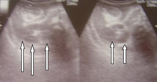
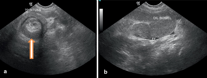
The ultrasonographic signs of intussusception include the target and pseudokidney signs as well as dilated bowel loops (Figs. 24.2 and 24.3).

Fig. 24.2
Abdominal ultrasound in a patient with intussusception showing pseudokidney sign

Fig. 24.3
a and b, Abdominal ultrasound showing the target sign (daunt sign). Note the dilated bowel loops in b
Stay updated, free articles. Join our Telegram channel

Full access? Get Clinical Tree


