Fig. 25.1
a and b, Diagrammatic representation of intestinal volvulus secondary to malrotation. The intestines twist in a clockwise direction around the superior mesenteric artery

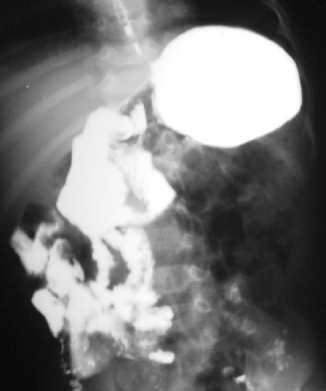
Fig. 25.2
Barium meal and follow-through in a patient with malrotation. Note that the duodenojejunal junction is lying on the right side of the spine and most of the small intestines are lying on the right side
It involves anomalies of intestinal fixation as well.
This can occur at a wide range of locations and leads to acute and chronic presentations.
The most common type found in pediatric patients is incomplete rotation predisposing to midgut volvulus, which can result in short-bowel syndrome or even death.
In 1936, William E. Ladd wrote the classic article on treatment of malrotation, and his surgical approach (Ladd procedure) remains the cornerstone of practice today.
The incidence of intestinal malrotation is one in 500 live births.
The male-to-female ratio is 2:1.
In infants, the mortality rate ranges from 2 to 24 %.
As many as 40 % of patients with malrotation present within the 1st week of life.
This condition is diagnosed in 50 % of patients by age 1 month and is diagnosed in 75 % by age 1 year. The remaining 25 % of patients present after age 1 year and into late adulthood; many are recognized intraoperatively during other procedures or at autopsy.
Embryology
Malrotation represents a spectrum of anomalies, and understanding the normal embryology of intestinal rotation is important in this regard. This happens in stages and the superior mesenteric artery forms the axis of rotation around which two loops rotate. The duodenojejunal loop begins to rotate superior to the superior mesenteric artery, and the cecocolic loop begins to rotate inferior to the superior mesenteric artery. Both loops make a total of 270°.
Stage I:
Occurs between the 5–10 weeks of gestation.
The intestines herniate into the base of the umbilical cord.
The duodenojejunal loop begins superior to the superior mesenteric artery at a 90° position and rotates 180° in a counterclockwise direction.
At 180°, the loop lies to the right of the superior mesenteric artery and by 270°; it lies beneath the superior mesenteric artery.
The cecocolic loop begins beneath the superior mesenteric artery at 270°.
It rotates 90° in a counterclockwise direction and lies to the left of the superior mesenteric artery at a 0° position.
Both loops maintain these positions until the bowel returns to the abdominal cavity.
Also during this stage, the midgut lengthens along the superior mesenteric artery, and, as rotation continues, a very broad pedicle is formed at the base of the mesentery. This broad base protects against midgut volvulus.
Stage II:
Occurs at 10 weeks’ gestation, the period when the bowel returns to the abdominal cavity.
As the bowel returns to the abdominal cavity, the duodenojejunal loop rotates an additional 90° to lie left of the superior mesenteric artery, the 0° position.
The cecocolic loop turns 180° more as it reenters the abdominal cavity. This turn places it to the right of the superior mesenteric artery, a 180° position.
Stage III:
This stage lasts from 11 weeks’ gestation until term.
During this stage, the cecum descends to the right lower quadrant.
Fixation of the mesenteries occurs at this stage.
Nonrotation:
Results from arrest in development at stage I.
The duodenojejunal junction does not lie inferior and to the left of the superior mesenteric artery, and the cecum does not lie in the right lower quadrant.
The mesentery has a narrow base.
This narrow base is prone to clockwise twisting leading to midgut volvulus.
The width of the base of the mesentery is different in each patient, and not every patient develops midgut volvulus.
Incomplete rotation :
Results from arrest in development at Stage II.
Most likely to result in duodenal obstruction.
Typically, the peritoneal bands running from the misplaced cecum to the mesentery compress the third portion of the duodenum leading to extrinsic duodenal obstruction.
Depending on how much rotation was completed prior to arrest, the mesenteric base may be narrow and, again, midgut volvulus can occur.
Internal herniations may also occur with incomplete rotation if the duodenojejunal loop does not rotate but the cecocolic loop does rotate. This may trap most of the small bowel in the mesentery of the large bowel, creating a right mesocolic (paraduodenal) hernia.
Incomplete fixation :
Failure of the mesentery of the right and left colon and the duodenum to become fixed retroperitoneally leads to the formation of potential hernial pouches.
If the descending mesocolon between the inferior mesenteric vein and the posterior parietal attachment remains unfixed, the small intestine may push out through the unsupported area as it migrates to the left upper quadrant. This creates a left mesocolic hernia with possible entrapment and strangulation of the bowel.
If the cecum remains unfixed, volvulus of the terminal ileum, cecum, and proximal ascending colon may occur. Malrotation and associated conditions
Intestinal malrotation is seen in association with the following conditions:
Gastroschisis, omphalocele, congenital diaphragmatic hernia, duodenal atresia, jejunoileal atresia, Hirschsprung’s disease, gastroesophageal reflux, intussusception , persistent cloaca, anorectal malformations, and extrahepatic anomalies.
Clinical Features
The clinical features of malrotation are variable depending on the age of the patient as well as the type and degree of malrotation. The presentations of malrotation are divided as follows:
Acute midgut volvulus:
This is the most serious presentation of malrotation.
Commonly seen in those < 1 year of age.
It is characterized by sudden onset of bilious vomiting and commonly abdominal distension.
Stay updated, free articles. Join our Telegram channel

Full access? Get Clinical Tree


