Chapter 33 Infections of the genital tract
VULVOVAGINAL INFECTIONS
The skin of the vulva, in common with other parts of the skin that are liable to friction and chafing, may be infected by common skin pathogens. Boils may occur, particularly when the standard of hygiene is low. Multiple vulval ulcers occur occasionally, particularly in debilitated women, and are due to staphylococcal infection. The ulcers are shallow, with a grey, discharging base and surrounding oedema. The vulva is extremely tender. Treatment consists of antibiotics and 1% chlorhexidine cream if this can be applied without causing much pain.
Genital herpes
Herpetic infection of the genital tract is becoming increasingly common. The virus, herpes simplex virus (HSV), exists in two forms, HSV1 and HSV2. Type specific HSV1 usually causes oral cold sores, but in up to 40% of cases is the cause of genital herpes. In the remainder HSV2 causes genital herpes. Serological testing shows that between 10 and 40% of adults have been infected at some time, but the infection was symptomatic in less than a quarter.
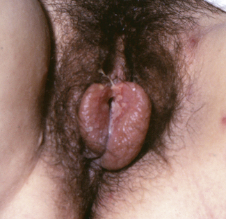
The first clinical attack of genital herpes is usually worse than the recurrences. It follows sexual contact with an infected person who was either symptomatically or asymptomatically shedding the virus. The inner surfaces of the labia majora are most likely to be infected. After a short period of itching or burning, small crops of painful, reddish lumps appear which become blisters within 24 hours. The blisters ulcerate rapidly to form multiple shallow, painful ulcers (Fig. 33.1). The surrounding tissues become oedematous and secondary bacterial infection may occur, aggravating the oedema and pain. Micturition may be very painful. Over 5 days the ulcers crust over and heal slowly, the healing being complete in 7–12 days after the appearance of the blisters for a primary infection, and less for recurrences. During this time, and intermittently, the virus is shed from the infected area and in vaginal secretions. The virus also enters the sensory nerves supplying the affected area, and tracks to lie in the dorsal root ganglion. It may lie dormant for the rest of the person’s life, or may be reactivated and track back along the nerves to cause a new attack of herpes. Second and subsequent attacks are less severe, but can cause considerable discomfort and affect relationships.
In 30% of infected women a single recurrence occurs, and between 2 and 5% have recurrent attacks, sometimes more than six times a year. As time passes the attacks (clinical and asymptomatic) become less frequent and may cease. In most cases the cause of the recurrence is not known, but recurrences are more common in the luteal phase of the menstrual cycle, if the woman has other sexually transmitted infections, or if she is emotionally stressed.
Intermittent asymptomatic shedding and atypical unrecognized lesions explain the unrecognized transmission to sexual partners. As HSV2 has a tropism for the genital area (HSV1 for the oral area) clinical recurrences are more frequent if the genital infection is HSV2 rather than HSV1.
Diagnosis
To make a definitive diagnosis of genital herpes, the blisters should be pricked to obtain vesicular fluid and the ulcers rubbed with a cotton tipped-bud to obtain virus-infected cells, and the swab sent in a virus transport medium for culture. Alternatively (particularly for older lesions) testing by polymerase chain reaction (PCR) is more sensitive. A full STI screen should be completed (Box 33.1).
Treatment
During an attack a woman should wash her hands after touching the infected area. If she is unable to pass urine because of pain or retention, a suprapubic catheter may need to be inserted. Local applications of ice or anaesthetic jelly provide some relief.
The antiviral drugs aciclovir, famciclovir and valaciclovir reduce the duration and severity of the initial and recurrent attacks, and shorten the time of viral shedding if given early in the attack. If a woman has five or more attacks each year, or the outbreaks are particularly severe, long-lasting or interfere with her psychosocial functioning, the drugs can be given daily as suppressive treatment for 6–12 months or longer.
Women who have recurrent genital herpes are often more concerned about the psychosocial sequelae of the disease, rather than the physical symptoms, and may need to be counselled.
Genital warts (condylomata acuminata)
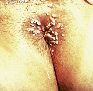
Genital warts are caused by types 6 and 11 of the human papilloma virus (HPV), usually transmitted sexually. Vulval infections are the most common, although the virus may spread to infect the vagina, the perineum (Fig. 33.2) and occasionally the cervix.
Vulval warts usually present as cauliflower growths of varying sizes, but may be clinically undetectable. It has been estimated that 5–10% of sexually active adults are infected annually, and that 30% have evidence of previous infection with other HPV genotypes.
In most cases the warts are symptomless, but some women complain of vulval discomfort, including itching. If the warts involve the vaginal entrance or the vagina, the woman may complain of dyspareunia.
The importance of HPV infections is that they are surrogates for the exposure to and carriage of the oncogenic HPV types 16 and 18, which are the cause of cervical carcinoma. The risk of this occurring from vulval warts is small. This is discussed further in Chapter 37.
Treatment
Genital warts, if not too exuberant, may be treated with podophyllotoxin (twice daily for 3 days, repeated if needed after 4 days) applied by the woman herself, after trimming away any vulval hair around the wart; or the medical practitioner may apply 20% podophyllin in benzoin tincture to the wart without touching the surrounding skin, to avoid skin burns and ulceration. The mixture is allowed to dry and the woman washes it off 6 hours later. This is repeated once weekly, if necessary. This should not be used in pregnancy. Larger condylomata on the cervix may respond to the application of trichloroacetic acid. Imiquimod cream 5% can be applied before bedtime so that it is left on the skin for 6–10 hours, three times weekly.
Large warts, or warts that fail to respond to medical treatment, are treated by diathermy or by laser. The procedure is painful and anaesthesia is needed. Postoperatively, pain requires analgesics. A problem that has recently been reported is that neither diathermy nor laser may cure the woman, as the virus may have infected neighbouring normal cells and warts commonly recur. Because of the concern that HPV infection with types 16 and 18 may be involved with the development of cervical carcinoma, a woman who has vulval warts should have regular Pap smears.
Syphilitic vulval ulcer
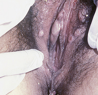
Syphilis is caused by invasion of the tissues by Treponema pallidum and is sexually transmitted. The primary lesion, which is often unrecognized, is a small papule that appears at the site of inoculation, usually 14–28 days after the person is infected. In women, the usual site of infection is one of the labia majora, but the cervix may be infected instead. The papule rapidly enlarges to form an oval lesion of variable size, the centre of which becomes eroded and granulomatous. The edges of the eroded area are sharp, and outside this a thickened, indurated zone occurs, hence the name for the lesion – hard chancre. The chancre is painless and may be ignored by the woman or considered a small sore of no consequence, but as it is teeming with treponemas it is highly infectious. The primary lesion disappears in 21 days or so. Secondary lesions, which include mucous patches and condylomata lata (Fig. 33.3), appear 5–6 weeks later.
Diagnosis
The diagnosis is confirmed by examining exudate expressed from the ulcer or mucous patches, through a microscope under dark-ground illumination. To obtain an accurate diagnosis the chancre is first cleaned with a swab, and then, if necessary, its edge and base are scarified with a scalpel so that exudate appears before the specimen is taken. The tests should be interpreted in conjunction with specialist sexual health physician or clinical microbiologist. A full STI screen should be ordered.
Infection of Bartholin’s gland (Bartholinitis)
The infection is usually due to Escherichia coli or staphylococci, but may follow Neisseria gonorrhoeae or Clostridium trachomatis infection. In the acute stage both the duct and the gland are involved. If it is not treated the infection may subside or a Bartholin’s abscess may form. Occasionally the gland becomes chronically enlarged following an inflammatory conglutination of the duct epithelium, to form a Bartholin’s cyst.
Diagnosis
In acute bartholinitis the woman complains of acute discomfort in the region of the gland, and a reddened, tender swelling appears beneath the posterior part of the labium majus.
Treatment
Treatment consists of excluding sexually transmitted infections (STIs) and prescribing analgesics and a broad-spectrum antibiotic. If an abscess has formed it should be marsupialized. An elliptical piece of the vagina and the abscess wall, just inside the hymen, is removed. The vaginal and abscess walls are sutured to maintain patency and a small drain is inserted.
Vaginal discharges and infections
The vagina of a woman in the reproductive years is lined by a layer of stratified epithelium, 10–30 cells thick. As is described on page 333, the superficial cells of the vagina are shed constantly into the vaginal cavity and release glycogen, which is acted on by Döderlein’s bacilli (lactobacilli) to produce lactic acid and hydrogen peroxide (natural defences against vaginal infection) and to maintain the vaginal pH between 3.5 and 4.5.
The vagina harbours large numbers of bacteria, which constitute its normal flora (Table 33.1). However, a significant number of women harbour potential pathogens such as E. coli, anaerobes, Candida spp., and Gardnerella vaginalis (bacterial vaginosis). They produce symptoms in only a few women.
Table 33.1 Normal vaginal flora

Stay updated, free articles. Join our Telegram channel

Full access? Get Clinical Tree

