Fig. 58.1
Histological picture showing features of Hodgkin’s lymphoma including the multinucleated Reed–Sternberg cells
Hodgkin’s lymphoma occurs at two peaks: the first in young adulthood (age 15–35) and the second in those over 55 years.
Hodgkin’s lymphoma may be treated with radiation therapy, chemotherapy, or hematopoietic stem cell transplantation. The choice of treatment depends on the age and sex of the patient and the stage, bulk, and histological subtype of the disease.
Patients with a history of infectious mononucleosis due to Epstein–Barr virus may have an increased risk of Hodgkin’s lymphoma, but the precise contribution of Epstein–Barr virus remains largely unknown.
Classification
Classical Hodgkin’s lymphoma (excluding nodular lymphocyte predominant Hodgkin’s lymphoma) can be subclassified into four pathologic subtypes based upon Reed–Sternberg cell morphology and the composition of the reactive cell infiltrate seen in the lymph node biopsy specimen.
Nodular sclerosing Hodgkin’s lymphoma :
The most common subtype.
It is composed of large tumor nodules showing scattered lacunar classical Reed–Sternberg cells set in a background of reactive lymphocytes, eosinophils, and plasma cells with varying degrees of collagen fibrosis/sclerosis.
Mixed-cellularity Hodgkin’s lymphoma :
It is the second most common subtype.
It is composed of numerous classic Reed–Sternberg cells admixed with numerous inflammatory cells including lymphocytes, histiocytes, eosinophils, and plasma cells without sclerosis.
This type is most often associated with Epstein–Barr virus infection.
Lymphocytic predominance Hodgkin’s lymphoma :
This is a rare subtype.
Shows many features which may cause diagnostic confusion with nodular lymphocyte predominant B cell non-Hodgkin’s lymphoma (NHL) .
This subtype has the most favorable prognosis.
Lymphocyte-depleted Hodgkin’s lymphoma :
This is a rare subtype.
Composed of large numbers of often pleomorphic Reed–Sternberg cells with only few reactive lymphocytes.
It may easily be confused with diffuse large-cell lymphoma.
Many cases previously classified within this category would now be reclassified under anaplastic large cell lymphoma.
Nodular lymphocyte predominant Hodgkin’s lymphoma expresses CD20, and is not currently considered a form of classical Hodgkin’s.
Staging
The staging is the same for both Hodgkin’s as well as NHLs .
Lymphangiograms or laparotomies are very rarely performed, having been supplanted by improvements in imaging with the computed tomography (CT) scan and positron emission tomography (PET) scan.
The Ann Arbor staging classification :
Stage I: Involvement of a single lymph node region (I) (mostly the cervical region) or single extra lymphatic site (Ie).
Stage II: Involvement of two or more lymph node regions on the same side of the diaphragm (II) or of one lymph node region and a contiguous extra lymphatic site (IIe).
Stage III: Involvement of lymph node regions on both sides of the diaphragm, which may include the spleen (IIIs) and/or limited contiguous extra lymphatic organ or site (IIIe, IIIes).
Stage IV: Disseminated involvement of one or more extra lymphatic organs.
The absence of systemic symptoms is signified by adding “A” to the stage; the presence of systemic symptoms is signified by adding “B” to the stage.
For localized extra nodal extension from mass of nodes that does not advance the stage, subscript “E” is added.
Splenic involvement is signified by adding “S” to the stage.
Clinical Features
Patients with Hodgkin’s lymphoma may present with the following symptoms :
Lymph nodes :
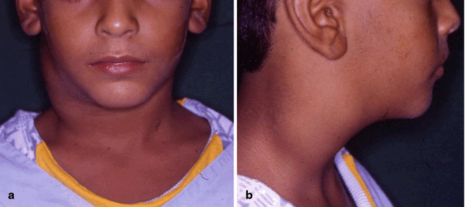
Painless enlargement of one or more lymph nodes (Fig. 58.2).

Fig. 58.2
Clinical photograph showing enlarged cervical lymph nodes in a child with Hodgkin’s lymphoma
The nodes may feel rubbery when examined.
The cervical and supraclavicular lymph nodes are most frequently involved (80–90 %).
The lymph nodes of the chest are often affected, and these may be noticed on a chest radiograph.
Itching.
Night sweats.
Weight loss.
Splenomegaly (30 %).
Hepatomegaly (5 %).
Back pain.
Red-colored patches on the skin, easy bleeding, and petechiae due to low platelet count.
Systemic symptoms: About one-third of patients with Hodgkin’s disease may also present with systemic symptoms, including low-grade fever; night sweats; unexplained weight loss, pruritus due to increased levels of eosinophils in the bloodstream; or fatigue.
Stay updated, free articles. Join our Telegram channel

Full access? Get Clinical Tree


