Fig. 6.1
a and b Clinical photographs showing extensive facial hemangiomas

Fig. 6.2
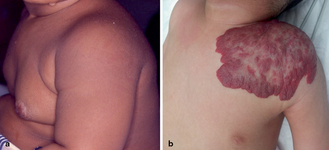
Clinical photograph showing hemangioma affecting the nipple (a) and anterior chest wall (b)

Fig. 6.3
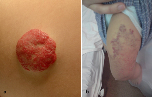
Clinical photographs showing hemangioma at the back (a) and lower limb (b)

Fig. 6.4
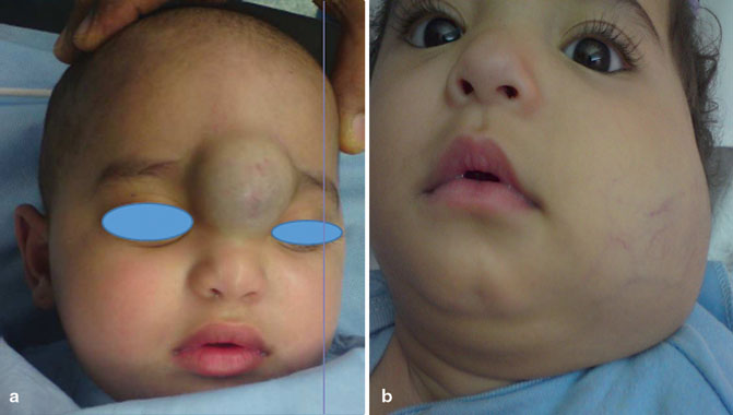
a and b Clinical photographs showing facial hemangiomas causing disfigurement
The cause of hemangioma is currently unknown; however, several studies have suggested the importance of estrogen in hemangioma proliferation.
Most hemangiomas are easily diagnosed without any additional diagnostic tests. Deeper hemangiomas or questionable superficial lesions, however, may require imaging studies to confirm the diagnosis and to evaluate their extent. Ultrasound (US) and magnetic resonance imaging (MRI) are valuable in their assessment.
Complications
The vast majority of hemangiomas are benign tumors and not associated with complications.
Hemangiomas may however cause several complications:
1.

Fig. 6.5
Clinical photograph showing an ulcerating hemangioma of the hand

Fig. 6.6
a – c Clinical photographs showing ulcerating hemangiomas
2.
It can cause bleeding.
3.
If a hemangioma develops in the larynx, breathing can be compromised.
4.
A hemangioma can grow and block one of the eyes, causing an occlusion amblyopia (Fig. 6.7).

Fig. 6.7
A clinical photograph showing hemangioma involving the lower eye lid. Further growth of the hemangioma may lead to lockage of the eye
5.
Very rarely, extremely large hemangiomas can cause high-output cardiac failure due to the amount of blood that must be pumped to all the hemangiomas blood vessels.
6.
Hemangiomas adjacent to bone can also cause erosion of the bone.
7.
Psychosocial complications might occur to the patient and family from large hemangiomas on the face (Fig. 6.8).

Fig. 6.8
a and b Clinical photographs showing facial hemangiomas that may cause psychosocial complications
8.
Children with large segmental hemangiomas of the head and neck can be associated with a disorder called PHACES syndrome (posterior fossa malformations, hemangiomas, arterial anomalies, cardiac defects, eye abnormalities, sternal cleft and supraumbilical raphe syndrome) .
9.
Rarely, large hemangiomas may cause Kasabach–Merritt syndrome (thrombocytopenia and hemangioma).
Treatment
In general, most hemangiomas disappear without treatment, leaving minimal or no visible marks.
Stay updated, free articles. Join our Telegram channel

Full access? Get Clinical Tree


