Case notes
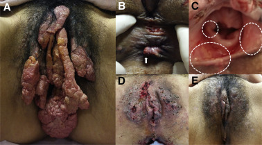
A 35-year-old woman who denied any previous sexual activity presented with an extremely large vulvar mass that had slowly grown for 25 years. The patient reported vulvar discomfort from the mass effect but no itching or pain. The vulvar mass consisted of 9 yellowish flesh-colored, cauliflower-shaped lumps, 115 × 90 mm diameter in total ( Figure 1 , A). The presentation initially suggested genital warts, so a human papillomavirus (HPV) DNA test with biopsy was performed. The results from polymerase chain reaction for both 9 types of low-risk and 19 types of high-risk HPV were all negative. The pathological diagnosis for the biopsy specimen was verruciform xanthoma.
Clinically, xanthoma masses usually occur in the oral mucosa or eyelid ; however, no evidence of xanthoma was found in the patient’s oral cavity or eyes. The patient had no medical problems, including dyslipidemia, and had no surgical history. Her immune status was normal and she had a normal white blood cell count and serum C-reactive protein level and had no history of recurrent infection. On her cheeks, there were scar bumps that we originally thought were caused by acne. However, considering the final diagnosis of xanthoma of the vulva, it is possible that these scars were xanthomatoses (we did not biopsy one).
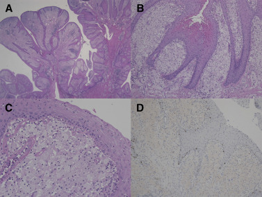
Under general anesthesia, we excised all the lumps using a scalpel and electrocauterization of the numerous xanthoma papules was performed. Hemostasis was further achieved by suturing the base of the lesions. A small xanthoma of anal canal ( Figure 1 , B) and multiple yellowish patches of mucosal xanthoma on the hymen and vestibule were noted ( Figure 1 , C). The patient performed dressing changes daily, until wound healing was complete about 3 weeks later. The immediate postoperative state and 2-month follow-up were grossly free of disease ( Figure 1 , D and E). The pathological examination revealed characteristic foam cells ( Figure 2 ) and magnetic resonance imaging showed a conglomerated vulvar mass ( Figure 3 ).
Stay updated, free articles. Join our Telegram channel

Full access? Get Clinical Tree


