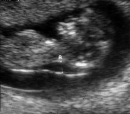
7 Genetic Screening and Obstetric Procedures Aria Koifman and Arnon Wiznitzer Maternal age of 35 years and over is the single most important risk factor for having a child with trisomy 21 (Down syndrome). In the past decade, amniocentesis and chorionic villous sampling (CVS) were introduced as methods of diagnosing Down syndrome for women who would be over the age of 35 years at time of delivery. Since both of these diagnostic techniques, which will be discussed later in this chapter, are quite invasive, noninvasive screening test were developed in the early 1980s and have since evolved. Today, several noninvasive screening strategies are available to clinicians. This chapter, which supplements Chapter 5, discusses the relative advantages and disadvantages of these common screening tests and their applications in common clinical practice. α-Fetoprotein: This is a major plasma protein, which is produced by the yolk sac and the liver during fetal life. Aneuploidy: This term refers to an abnormal number of chromosomes. Syndromes caused by an extra or missing chromosome are among the most widely recognized genetic disorders in humans. Dimeric inhibin A: This is a glycoprotein of placental origin, similar to human chorionic gonadotropin. Levels of this glycoprotein in maternal serum remain relatively constant through the 15th to 18th weeks of pregnancy. Maternal serum levels of dimeric inhibin A are twice as high in pregnancies affected by Down syndrome than in unaffected pregnancies. Human chorionic gonadotropin: This glycoprotein hormone is produced in pregnancy, by the developing embryo soon after conception and later by the placenta. Its role is to prevent the disintegration of the corpus luteum of the ovary and thereby maintain progesterone production that is critical for the maintenance of a healthy pregnancy in humans. Nuchal translucency: This term is used to describe the area at the back of the neck (nuchal region) of the developing baby as assessed by ultrasound scanning. Measurement of the nuchal translucency is performed in the first trimester (between 11 weeks and 13 weeks, 6 days). Measurements of greater than 3.0 mm may warrant further investigation. Pregnancy associated plasma protein A: This is a large, zinc-binding protein that acts as an enzyme, specifically a metallopeptidase. Women with low blood levels of this protein at 8–14 weeks of gestation have an increased risk of intrauterine growth restriction, trisomy 21, premature delivery, pre-eclampsia, and stillbirth. Trisomy: This is a form of aneuploidy with the presence of three copies, instead of the normal two, of a particular chromosome. Down syndrome, in which affected individuals have an extra copy of chromosome 21, is called trisomy 21. Unconjugted estriol: Estriol is a steroid hormone derived from cholesterol and is the major estrogen produced during pregnancy. It is only produced in significant amounts during pregnancy as it is made by the fetal liver and the placenta. Although current screening strategies offer high sensitivity and specificity rates, patients should be informed that a screening test provides an individual risk assessment but is not diagnostic for chromosomal abnormalities. The main disadvantage of screening tests is that not all affected fetuses will be detected. A screen is considered positive when a value for one or more of the screened disorders falls above a designated risk cutof. A risk cutof might be chosen based upon the desired detection rate, false-positive rate, or a combination of both. The results in the screening tests are expressed (aside from their proper measuring units for each marker) in multiples of the median (MoM). Each marker result, including both biochemistry and nuchal translucency measurements, can be expressed in MoM. This allows for direct comparison of results between laboratories. Historically, a biochemical marker in maternal serum, combined with maternal age, has been the main screening method. As ultrasound imaging became a common practice in obstetrics, it was only natural to use it in aneuploidy screening. Combination of the methods is the basis for current screening strategies. In cases identified as “screen positive,” proper counseling should be provided in order to deliver the information in a clear and accurate manner. One early study reported that increased nuchal translucency (NT), which can be visualized by ultrasound scanning at 11–14 weeks of gestation (Fig. 7.1), is associated with trisomy 21. Unfortunately, the detection rate, which was validated by several studies, was only 65–71%, with a false-positive rate of about 5–8%. Only with large-scale studies, when the option of using NT in a quantitative manner (i. e., with MoM) became available, could NT be combined with biochemical markers to improve detection rates for trisomy 21. Fig. 7.1 Ultrasound scan at 11 weeks’ gestation showing a thickened nuchal translucency (A) of 3 5 mm NT has proven to be useful not only in aneuploidy screening, but also for fetal cardiac malformations, diaphragmatic hernia, and some rare single-gene disorders as well. An NT value above the 95th percentile warrants a detailed ultrasonographic anatomy scan, and in many centers even fetal echocardiography is employed in such cases. The most widely studied markers in the first trimester of pregnancy are free beta human chorionic gonadotropin (β-HCG), total β-HCG, and pregnancy associated plasma protein A (PAPP-A). Of these, β-HCG has been found to be useful in both the first and second trimesters and provides predictive information concerning pregnancy outcomes. The level of PAPP-A, a glycoprotein produced by the trophoblast, is reduced in Down syndrome and trisomy 18. This marker is only useful as a quantitative screen in the first trimester. Regarding detection rates, first-trimester biochemical markers alone are not superior to the second-trimester triple test (see below). However, as previously mentioned, PAPP-A in combination with NT provides detection rates of 82–87%. Around 10% of circulating estriol (E3) in the maternal compartment remains in the unconjugated form. Decreased levels of uconjugated estriol (uE3) have been seen in pregnancies affected with Down syndrome in the second trimester of pregnancy. Low levels of midtrimester uE3, alone or in association with other midtrimester markers, has been associated with adverse pregnancy outcomes.
Definitions
Screening Strategies
First Trimester Screening
Nuchal Translucency

Biochemical Markers
Second Trimester Screening Tests
Triple Test
Stay updated, free articles. Join our Telegram channel

Full access? Get Clinical Tree


