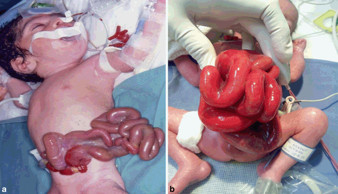
Fig. 36.1
A clinical photograph showing gastroschisis. Note the site of the herniated intestines to the right of the umbilicus
Omphalocele on the other hand is a congenital birth defect through the umbilical cord and the contents remain enclosed in a sac of visceral peritoneum. With omphalocele, the defect is usually much larger than in gastroschisis .
Gastroschisis is a relatively uncommon condition. It occurs in approximately 1 out of every 5000 live births. It has been reported that the incidence of gastroschisis has increased in recent years.
Gastroschisis is usually inherited in an autosomal recessive manner. It may begin as a sporadic mutation, can be associated with nongenetic congenital disorders, but has also been observed to be autosomal dominant.
During the 4th week of development, the lateral body folds move ventrally and fuse in the midline to form the anterior body wall. Incomplete fusion results in a defect that allows abdominal viscera to protrude through the abdominal wall. The bowel typically herniates through a defect to the right of the umbilicus.
The malformation is slightly more frequent in males than females.
The frequency of gastroschisis is associated with young maternal age and low number of gestations.
Etiology
The exact etiology of gastroschisis is not known.
It is associated with younger maternal age and almost never occurs in mothers over 30 years of age.
The following factors have been incriminated as possible etiological factors:
The use of salicylates
Maternal cigarette smoking
Maternal alcohol and drug use
Several embryological hypotheses have been proposed as contributing factors for gastroschisis. These include:
1.
Failure of mesoderm to form in the body wall
2.
Rupture of the amnion around the umbilical ring with subsequent herniation of bowel
3.
Abnormal involution of the right umbilical vein leading to weakening of the body wall and gut herniation
4.
Disruption of the right vitelline (yolk sac) artery with subsequent body wall damage and gut herniation
5.
Abnormal folding of the body wall which results in a ventral body wall defect through which the gut herniates
6.
Failure to incorporate the yolk sac and related vitelline structures into the yolk sac
Diagnosis
The diagnosis of gastroschisis is commonly made antenatally by a routine ultrasound examination.
Rarely, polyhydramnios may prompt an antenatal ultrasound examination.
The herniated bowel in gastroschisis is bathed by amniotic fluid and both maternal serum and amniotic fluid alpha-fetoprotein (AFP) levels are elevated. This should be evaluated by an abdominal ultrasound.
Maternal abdominal ultrasound usually shows the herniated bowel and perhaps the liver floating in the amniotic fluid.
Plans should be made for careful delivery and immediate management after birth.
Ultrasound may also reveal intrauterine growth retardation which occurs in 38–77 % of fetuses with gastroschisis .
This is usually secondary to nutrient loss through exposed intestines.
Approximately 48 % of infants with gastroschisis are small for their gestational age.
Examination
Clinically, the appearance of bowel may range from almost normal-to-thick-walled inflamed intestines forming a mass (Fig. 36.2).


