Rationale for Use in Endometrial Cancer
The extent to which the para-aortic nodes should be removed remains under some debate; it is our opinion that adequate assessment requires dissection up to the level of the renal veins rather than to the inferior mesenteric artery (IMA). Extension of the para-aortic lymphadenectomy has the potential to double the count of lymph nodes available for evaluation [16] and may detect metastasis in patients without nodal disease below the inferior mesenteric artery. In a study of 281 patients undergoing lymphadenectomy for surgical staging of endometrial cancer, 67 % had para-aortic disease. Of those, 77 % had involvement of nodes above the inferior mesenteric artery. For patients with para-aortic nodal involvement, 46 % were documented to have positive nodes above the inferior mesenteric artery and negative ipsilateral nodes below [13].
However, performing an adequate para-aortic lymphadenectomy to the renal veins can be challenging, particularly in obese patients and when using minimally invasive surgical techniques. In the LAP2 trial conducted by the Gynecologic Oncology Group, comparing laparoscopic to open surgical staging for uterine cancer, risk of conversion to laparotomy increased with higher body mass index (BMI). 26 % of those assigned to laparoscopy required conversion to laparotomy to complete staging. The most common reason for conversion was poor visualization. Across all subgroups, comprising differences in age and metastatic disease, BMI remained an important risk factor for conversion, with an odds ratio of 1.11 for each 1-unit increase in BMI [17]. A study comparing laparoscopic to open surgery with planned pelvic and para-aortic lymph node dissection in obese endometrial cancer patients found similar lymph node counts in both groups (extent of para-aortic dissection was to the inferior mesenteric artery) but noted a 36 % conversion rate, with the most common reason reported as obesity. Again, successful laparoscopic lymphadenectomy was less likely with increasing BMI, particularly greater than 35 [18].
Indications and Technique
Extraperitoneal laparoscopic para-aortic lymphadenectomy may be appropriate for patients at high risk for lymphatic disease or for patients in whom visualization for infrarenal dissection may be challenging (obese, prior surgery, etc.). Using this technique, patients may be staged by vaginal hysterectomy and extraperitoneal pelvic/para-aortic lymphadenectomy. Silver and colleagues argue that the latter approach may arguably be the least invasive method of staging endometrial cancer, as it greatly limits intraperitoneal disruption and subsequent adhesion formation that may contribute to postoperative and post-radiation complications [19]. Contraindications to extraperitoneal laparoscopic para-aortic lymphadenectomy include a history of retroperitoneal surgery (e.g., nephrectomy) or contraindications to laparoscopy such as severe cardiopulmonary disease or closed-angle glaucoma.
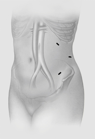
Prior to initiation of extraperitoneal laparoscopic para-aortic lymphadenectomy, it is important to assess the intra-abdominal cavity for metastatic disease, which may be performed via a transperitoneal umbilical port. If no metastatic disease is identified, a total of three ports are placed in the left flank, with proper placement critical to the success of the procedure so as not to constrict the operative field or cause perforation of the peritoneum. An initial 2-cm incision is made two fingerbreadths medial and three to six fingerbreadths superior to the left anterior superior iliac spine. Fibers of the oblique and transversalis muscles are split bluntly until the peritoneum is identified. Blunt dissection is continued to develop the retroperitoneal space posteriorly until the left psoas muscle is palpated. A second incision is then made superior and inferior to the first, and a 10-mm trocar is inserted. The retroperitoneal space is insufflated, keeping initial pressures (10 mm Hg) and flow (3 L/min) low to minimize the risk of peritoneal perforation and possible subsequent pneumothorax and hypercarbia. The camera is inserted through this port and additional blunt dissection is performed through the initial incision until the psoas muscles are visualized. A 5-mm trocar is placed under direct visualization further superior and anterior to the second, and a 10-mm port is placed in the initial incision. Port placement is outlined in Fig. 19.2. Pressure may be gradually increased if exposure is inadequate. However, pressures greater than 15 mm Hg should be avoided.
Insufflation pressure generally allows passive retraction of the left ureter and gonadal vessels anteriorly out of the field. Dissection is continued medially with identification of the left common iliac artery and aorta. Following the left gonadal vein into the left renal vein superiorly identifies the superior most limits of the dissection, and para-aortic nodes between the aortic bifurcation and left renal vein are then removed. Dissection is then continued medially to develop the space over the aorta to the inferior vena cava. The right para-aortic lymph nodes are then reflected from the underlying inferior vena cava, with insufflation pressure allowing them to be retracted anteriorly to the roof of the dissection. The inferior mesenteric artery is identified and preserved. Due to the approach from the left side, identification of the right ureter is unnecessary but it may usually be seen along the lateral aspect of the dissection. The nodes are then stripped from the anterior peritoneum. Once all nodal tissue has been removed, the lowermost trocar may be converted to an intraperitoneal port by advancing it through the peritoneum. Transperitoneal pelvic lymphadenectomy may then be performed with placement of additional transperitoneal ports. Use of this technique has been reported using a single-port approach, although this is not widely utilized [20].
Feasibility and Outcomes
As the technique was first described for para-aortic nodal evaluation in the setting of cervical cancer, many of the early studies regarding feasibility arise from that body of literature. In an early paper, Dargent first reported this technique in 44 patients with cervical cancer who underwent laparoscopic para-aortic lymphadenectomy, by either a transperitoneal, bilateral extraperitoneal, or left-sided extraperitoneal approach. Success rates for the respective methods were 78 %, 93 %, and 95 %, although comparison of techniques in this study is limited due to small numbers and the learning curve of the surgeons. Conversion to a transperitoneal approach due to peritoneal perforation occurred in 17 % of extraperitoneal attempts. The extraperitoneal approach was initially pursued because of the difficulty obtaining adequate retraction of bowel and visualization in order to safely perform lymphadenectomy up to the renal veins. The left extraperitoneal approach also yielded equivalent node counts with a shorter operative time [2]. In a larger study by the same group, 53 patients underwent attempted infrarenal para-aortic lymphadenectomy using a laparoscopic extraperitoneal approach, with an overall success rate of 96 % and average nodal count of 20.7. The average procedure time was approximately 126 min [1]. Other groups have reported similar outcomes [21, 22].
The Mayo Clinic experience with laparoscopic extraperitoneal para-aortic lymphadenectomy is one of only a few studies specifying results with regard to successful dissection up to the renal veins. Over 2 years, 38 patients underwent attempted para-aortic lymphadenectomy using the technique, with a 92 % success rate; the mean BMI was 33 and the remainder of the procedure was in most cases performed vaginally. Median operating time for the para-aortic lymphadenectomy portion of the case was 69 min, with an average of 16.5 nodes removed. While not statistically significant, there was a trend toward higher nodal counts for patients with a greater BMI (over 35). An average of 9.5 nodes was harvested above the inferior mesenteric artery. Indeed, in the most obese patient (BMI of 52), 21 of 34 lymph nodes were harvested above the IMA [23].
The technique is neither difficult to adapt nor arduous to disseminate to trainees. In a study reporting outcomes for the procedure when taught to gynecologic oncology fellows, after an average of 5 mentored cases as an assistant surgeon, 100 % of 22 planned operations were completed successfully, with an average nodal count of 14 and estimated blood loss of 40 mL. Operating times were understandably longer at a mean of 163 min, with an acceptably low complication rate of 6.2 % [24]. In a small study following inexperienced surgeons performing porcine para-aortic lymphadenectomy via either a transperitoneal or extraperitoneal route, the learning curves were similar for both approaches. Ten animals were required for each approach in order for the physician to perform the procedure effectively; one would expect that number to be lower for more experienced laparoscopic surgeons adapting the new technique [25].
Stay updated, free articles. Join our Telegram channel

Full access? Get Clinical Tree


