Epstein-Barr Virus Mononucleosis
W. Garrett Hunt and Michael T. Brady
The Epstein-Barr virus (EBV) is recognized as the major cause of heterophil-positive and heterophil-negative infectious mononucleosis. Manifestations of EBV infection are varied and range from asymptomatic infection to fulminant lymphoproliferative disease. The virus is associated with a number of malignancies, including African Burkitt lymphoma, nasopharyngeal carcinoma, Hodgkin disease, and a spectrum of posttransplant lymphoproliferative diseases. The specific role of EBV in each tumor is now defined, in a number of circumstances, to the level of specific cell type and receptors, intra-cellular pathways, gene expression, and cytokine production.
 EPIDEMIOLOGY
It is important to recognize that acute primary Epstein-Barr virus (EBV) infection is not synonymous with infectious mononucleosis. Most EBV infections acquired at any age, but particularly during childhood, are asymptomatic. Seroepidemiologic studies demonstrate that from 20% to 100% of children worldwide have antibodies to EBV by 6 years of age.9 In contrast, in the United States, only 40% to 50% of adolescents are seropositive,9-11 with higher socioeconomic groups being less likely to have evidence of prior infection. Seropositivity increases with age in all populations, so that almost all adults have serologic evidence of past EBV infection. Seroconversion is particularly high in college, where 10% to 15% of susceptible persons become infected each year. This group of EBV-naive adolescents in industrialized countries is susceptible to develop EBV-associated IM, much more common in the United States and Western Europe than in unindustrialized countries.
EBV is excreted in oropharyngeal secretions and is transmitted by contact with saliva via kissing or other mucosal contact with contaminated objects.12 Healthy seropositive individuals intermittently shed EBV into their oropharynx. Blood products or transplanted tissues can transmit EBV and are particularly problematic for seronegative immunocompromised transplant recipients. There is no evidence of urinary or fecal excretion. Transplacental transmission appears to be rare. Shedding of virus appears to be more frequent in immunosuppressed individuals, 60% of whom may excrete EBV at any one time. Because virus shedding is of a low titer in even immunocompromised patients, standard precautions are adequate for isolation of patients with acute or past EBV infections.12
The epidemiology of infectious mononucleosis is closely related to the age of primary EBV infection. In the United States, the incidence of infectious mononucleosis is approximately 50 per 100,000 persons per year, but in individuals 15 to 25 years old, the incidence doubles.13 Those areas where children are infected at an early age have the lowest incidence of the disease. Among susceptible adolescents and young adults, studies measuring both apparent and inapparent EBV infections indicate a clinical-to-subclinical ratio of 1:2 to 1:3.
 PATHOPHYSIOLOGY
Epstein-Barr virus (EBV) is a member of the family Herpesviridae (gamma herpesvirus), which contains linear double-stranded DNA surrounded by a protein capsid with 162 cap-somers in an icosahedral arrangement.1 The nucleocapsid is covered by a lipid-containing envelope derived from the nuclear membrane of the host cell. EBV causes lytic infection of human oropharyngeal and salivary cells, and latent infection of human and primate B lymphocytes and epithelium of the nasopharynx.2 This virus has been long recognized to be lymphotropic for B lymphocytes and to infect both oropharyngeal epithelial cells and myocytes, but it has also been recognized that it infects T lymphocytes, natural killer (NK) cells,3,4 and monocytes, which may serve as an early site for viral replication.5
Infection of lymphocytes with EBV can transform them into continuously growing lymphoblastoid cell lines containing circular genome as a plasmid. Once infected, transformed lymphoblastoid cells rarely continue to produce infectious virus in vitro, although EBV-induced antigens can be detected in the cells. The appearance of new antigens on the cell surface of EBV-infected cells is believed to be responsible for the cellular immune response to the virus and for the pathogenesis of the disease produced.6-8 The EBV receptor on epithelial cells and B lymphocytes is the CD21 molecule (formerly CR2), which is also the receptor for the C3d fragment of the third component of complement. The virus elicits both humoral and cellular immune responses.
Epstein-Barr virus (EBV) acquired by ingestion appears to first infect oropharyngeal epithelial cells. Subsequently, the virus infects susceptible B lymphocytes within the lymphoid tissue of the pharynx. During a 30- to 50-day incubation period, virus actively replicates and disseminates throughout the entire lymphoreticular system.14
Cell-mediated immune function is essential in the control of and recovery from EBV infection. In EBV infectious mononucleosis, the initial infection of B lymphocytes is followed by an extensive proliferation of suppressor cytoxic T lymphocytes (CD8+ positive). Associated with the increase in these cytotoxic and suppressor cytotoxic T cells is a concomitant decrease in the number of T-helper/inducer cells (CD4+-positive lymphocytes), resulting in an inversion of the CD4:CD8 ratio. Fatal infectious mononucleosis and lymphoproliferative disorders, in which B-cell lymphomas develop, have been identified in adults and children with such cell-mediated immune defects, and, in particular, in their natural killer (NK) cell activity. Groups identified as developing this progressive B-immunoblastic disorder include kidney, heart, and bone marrow transplant recipients and individuals with X-linked lymphoproliferative syndrome, severe combined immunodeficiency syndrome, AIDS, ataxia-telangiectasia, and certain autoimmune diseases.
More recently, EBV has been demonstrated to infect T cells or NK cells to cause unique systemic lymphoproliferative diseases such as EBV-associated hemophagocytic lymphohistiocytosis (EBV-HLH), as well as chronic active EBV (CAEBV) infection, clinical features of which are distinct from the tumor-forming diseases outlined previously.3,4 Systemic EBV-related lymphoproliferative disease (LPD) is now classified into B-cell LPD and T/NK-cell LPD. The former causes fulminant infectious mononucleosis, whereas the latter causes EBVHLH and CAEBV.
 CLINICAL MANIFESTATIONS
Infectious Mononucleosis
The incubation period of infectious mononucleosis syndrome is 30 to 50 days. The clinical syndrome of infectious mononucleosis is usually preceded by a 3- to 5-day prodrome of malaise, fatigue, headache, nausea, or abdominal pain.15 Over the next 7 to 20 days, sore throat and fever gradually increase. The triad of fever, sore throat, and posterior cervical adenopathy occurs in more than 80% of patients. Sore throat is often accompanied by evidence of moderate-to-severe pharyngitis, with marked tonsillar enlargement that may be covered with shaggy gray or white exudates. Fine petechiae may cover the uvula and soft palate during the initial week of illness. Throat cultures are positive for group A β-hemolytic streptococci in about 30% of patients, which may confuse the correct diagnosis of Epstein-Barr virus (EBV) mononucleosis.2,15,16 Fever is present in 85% to 95% of patients, from 39°C (102°F) up to 40.5°C (105°F), and on average lasts 10 days, but may persist for weeks.2
Adenopathy most often involves only the bilateral posterior cervical nodes but can involve any nodes. The nodes are affected singly or in groups (not necessarily symmetrically) and may be very large or small (the size of grapes); they are most often firm, discrete, and moderately tender to palpation. Splenomegaly, with the spleen palpable 2 to 3 cm below the costal margin beyond the neonatal period, occurs in about 50% of infections.17 Rupture is rare but can be a potentially fatal complication. Hepatomegaly occurs in 10% to 30% of patients, but less than 5% of patients develop jaundice. Serum aspartate aminotransferase (AST) and serum lactate dehydrogenase (LDH) are mildly elevated in the majority of patients and may persist for weeks to months. Chronic liver disease, however, does not typically result.
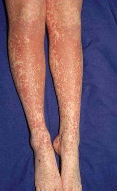
Other clinical findings include bilateral supraorbital edema and rashes. A blanching, erythematous, maculopapular exanthema occurs in about 5% to 15% of patients, but as many as 80% develop this rash if treated with ampicillin or other β-lactam antibiotics (Fig. 311-1). The same rash may occur with cytomegalovirus (CMV)-associated mononucleosis and so does not differentiate CMV- from EBV-associated mononucleosis. Urticarial, bullous, hemorrhagic, and scarlatiniform rashes, as well as the Gianotti-Crosti syndrome, are also associated with infectious mononucleosis.
Neurologic complications include aseptic meningitis, encephalitis, optic neuritis, GuillainBarré syndrome, transverse myelitis, Bell palsy, and, in numerous more recent epidemiologic studies, multiple sclerosis following EBV-associated infectious mononucleosis.18 An autoimmune hemolytic anemia occurs in 0.5% to 3% of infectious mononucleosis patients and is usually mediated by antibodies against the “i” antigen. A mild thrombocytopenia below 140,000 platelets/μL, but with absence of profound thrombocytopenia or bleeding, occurs in approximately 50% of patients. Granulocytopenia, or thrombocytopenia may occur during the acute illness or in the immediate recovery period. Respiratory and cardiac complications include interstitial pneumonia, laryngeal obstruction, pharyngeal edema, myocarditis, and pericarditis.
Stay updated, free articles. Join our Telegram channel

Full access? Get Clinical Tree


