Enterobiasis (Pinworm)
Richard A. Oberhelman
 EPIDEMIOLOGY
Enterobiasis is caused by the pinworm Enterobius vermicularis, a strictly human parasite infecting the gastrointestinal tract. Infection occurs worldwide, and clustering of cases in families is common.
In the United States, infection rates in young school children vary from 10% to 45%. Infection is unrelated to poor sanitary facilities or tropical climates. Young girls have pinworm more frequently than boys of the same age, and whites are more often infected than African Americans. Infection is most common between early fall and late spring, perhaps related to transmission in schools. For unknown reasons, some individuals seem to be predisposed or vulnerable to reinfection.
 PATHOPHYSIOLOGY
Infection occurs by ingestion of embryonated eggs excreted in the stool of infected persons and may occur by hand-to-mouth transmission or by oral contact with infected fomites, such as toys, bedding, or clothing. The eggs average 55 μm by 35 μm and appear flattened on one side and convex on the other. They are fully mature and infective 3 to 8 hours after being deposited, but at normal room temperature, less than 10% of eggs live for 48 hours. Ingested eggs with first-stage larvae hatch in the duodenum, and the larvae develop into adults in the cecum, where they mate. The gravid female detaches from the cecal mucosa and migrates down the large bowel, usually passing out the anus onto the perianal and perineal skin, leaving a trail of eggs on the surface of the skin. Yellow-white female adult pinworms measuring 8 to 13 mm may be seen emerging from the rectum of infected children, most often around 10 or 11 pm. In approximately 5% of patients, eggs are deposited in the bowel and may be found in feces. Generally, the worm dies after ovipositing is completed, so repeated infections are the result of autoinfection or reinfection from other environmental sources. There is no good evidence that retrograde infection occurs.1
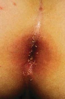
 CLINICAL MANIFESTATIONS
Pinworms rarely produce serious pathology, and many infections are asymptomatic. Perianal and perineal pruritus are the most common complaints.2 Although pruritus probably results from crawling worms, some patients with heavy pinworm infections and many worms in the rectum have little or no itching. Pruritus may provoke such severe scratching that local bleeding, secondary pyogenic infection, and lichenification can occur. Whether pinworms are a primary cause of appendicitis remains unsettled; most pathologists consider their presence in an acutely inflamed appendix to be incidental, although infections in the colon with associated gastrointestinal cramping and diarrhea have been reported. Vaginal infection in young girls is common and may be associated with vaginitis and discharge. Pinworms occasionally have been found in the fallopian tubes, resulting in intraabdominal ectopic migration and symptomatic granulomatous inflammation in the peritoneal cavity.
Stay updated, free articles. Join our Telegram channel

Full access? Get Clinical Tree


