Disorders of the Foot
Charles E. Johnston
A common referral to a pediatric orthopedist is for a foot deformity that may or may not be symptomatic. Conditions range from benign, self-resolving, perceived abnormalities involving the forefoot and toes, to more severe congenital and neuropathic deformities including clubfoot, congenital vertical talus, and cavus foot. Frequently, differentiation between a benign, resolving condition and a more severe pathologic deformity can be made by clinical examination and level of suspicion. A review of the common disorders, both benign and pathologic, will be presented to assist in the office evaluation of pediatric foot conditions.
CONGENITAL DEFORMITIES
 METATARSUS ADDUCTUS
Medial deviation of the forefoot of an infant, termed metatarsus adductus, is one of the most common pediatric foot conditions (Fig. 213-1). Intrauterine positioning—medial rotation of the foot across the fetal torso—is the presumed cause of this positional deformity.
When viewed from the plantar surface, the lateral border of the foot is curved and appears “bean shaped” (Fig. 213-1B). There may be additional medial deviation of the great toe and the appearance of a high arch.2 The deformity can be passively “corrected” if the examiner grasps the heel and maintains it in the neutral position, while abducting the forefoot. Metatarsus adductus is almost always bilateral, although some children will demonstrate a “windswept” position of the feet, with one foot internally rotated with metatarsus adductus, and the opposite externally rotated by the intrauterine position.
Many children will present at walking age with intoeing. Metatarsus adductus is but one reason the foot progression angle can be deviated medially—the most common being internal tibial torsion (ITT). Metatarsus adductus is distinguished from talipes equinovarus (clubfoot) by noting that the ankle and hindfoot have a normal range of dorsiflexion and plantarflexion, whereas a true clubfoot has a fixed hind-foot deformity (see discussion below). Rarely, the forefoot may be medially deviated in a foot that appears to have severe midfoot and hind-foot valgus, a foot that is commonly described as skewfoot (see discussion below).
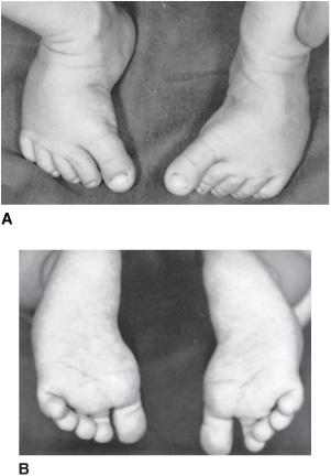
FIGURE 213-1. A: Dorsal view of bilateral metatarsus adductus. Note the medial deviation of all toes. (Reprinted with permission from Tachdjian’s Pediatric Orthopaedics, 4th Edition, edited by John A. Herring, Fig. 23-19A.) B: Plantar view. The lateral border of the foot is curved and “bean shaped.” (Reprinted with permission from Tachdjian’s Pediatric Orthopaedics, 4th Edition, edited by John A. Herring, Fig. 23-19B.)
Treatment of metatarsus adductus varies from observation to casting in more severe presentations.2,3 If the forefoot can be passively manipulated into a corrected position, then the parents can be instructed in passive stretching, and the “deformity” can be observed. Full resolution can be expected. Night splints or reverse-last shoes for children younger than 6 months can also be utilized, although there is little evidence that such orthotic treatment facilitates what would otherwise be spontaneous resolution. If the forefoot is not passively correctable, a short period of casting may be warranted. 
Surgical correction is reserved for the rare patient with a fixed adductus, and symptoms. The decision for operative treatment is controversial. Orthopedic consensus is that, even with moderate fixed residual adductus, adult function is still normal.2,3
 CALCANEOVALGUS FOOT
This postural deformity of infancy is characterized by a sometimes dramatic hyperdorsiflexion position, where the dorsum of the foot is plastered to the anterior surface of the tibia (Fig. 213-2). Plantarflexion is limited, and the condition may seem severe if the contractures of the dorsal ankle structures do not allow early manipulative relief  .
.
The foot may also be deviated laterally, the “valgus” portion of the deformity, preventing inversion of the foot. The key is to determine whether the heel is also in dorsiflexion. If the heel has descended and moves appropriately upward when the forefoot is plantarflexed, one may conclude that this is a postural, in utero deformity, resulting as a “packaging” defect, which will resolve with time and manipulative stretching.4,5 On the other hand, if the heel has not descended with the hyperdorsiflexed forefoot or does not move appropriately when the forefoot is raised and lowered, then a congenital vertical talus must be ruled out. The latter is usually characterized by the foot having a “rocker bottom” appearance, due to the fixed equinus of the heel (see discussion below). Hip dysplasia must be ruled out in the newborn with a hyperdorsiflexed foot, because the same abnormal in utero fetal position responsible for the calcaneovalgus foot can also cause hip dysplasia.5
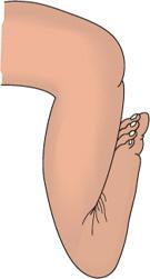
FIGURE 213-2. Hyperdorsiflexed position of a calcaneovalgus foot.
The hyperdorsiflexed foot can be treated by gentle manipulation and stretching, pushing the foot into plantarflexion (eFig. 213.2 ). Such stretching exercises can be taught to the parents. Inversion of the foot may also be carried out as a corrective maneuver. Typical calcaneovalgus foot will resolve spontaneously in a period of 3 to 6 months, although occasionally a more severe case may be referred to the orthopedist for either splinting or casting. Parents should be educated as to the benign nature of this deformity, but they should also be warned that there may be a flatfoot appearance when ambulation begins.
 FLEXIBLE FLATFOOT (PES PLANOVALGUS)
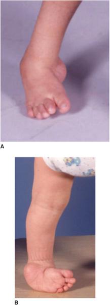
Flatfoot is probably the most common “deformity” evaluated by pediatric orthopedists. Whether it constitutes an actual deformity is, in fact, questionable.7 In general, the weight of the child’s body in the presence of lax ligaments in the foot flattens the normal arch. In the infant and pretoddler child, a “fat” foot should not be mistaken for a flatfoot, as the arches of infants and young children are often obscured by subcutaneous fat.8 Finally, a lower arch is a normal variant.
The appearance of a flatfoot is well known—the heel is everted with a loss of height, and the talar head and navicular appear to rest on the floor medially. The medial column of the foot may appear longer than the lateral column. When viewed from behind, the examiner may note that all five toes are visible to the lateral side of the tibia (called “too many toes” sign). However, while observing the child from behind, the examiner may request that the child go up on tiptoes, at which time in the flexible flatfoot, the longitudinal arch will reconstitute, and the heels will invert (eFig. 213.3  ). This simple maneuver basically rules out any significant congenital foot pathology.
The differential diagnosis of flatfoot includes conditions such as congenital vertical talus, in which the heel is in equinus and will not correct, and tarsal coalition, in which there is marked restriction of subtalar and midfoot movement, most easily detected by the tiptoes test just described. Tarsal coalition usually presents in older children, typically because of pain.
A decreased longitudinal arch has been described in as much as a quarter of the adult population, and thus must be considered a normal variant. This, however, does not remove the possibility that the patient may have symptoms, which are almost always associated with a contracture of the triceps surae.9 Should heelcord contracture be suspected, the child should be asked to walk on his or her heels, which will be difficult with a significant contracture.
Treatment of flexible pes planovalgus depends on the presence or absence of symptoms. Asymptomatic flatfeet require no treatment, with education and reassurance being given to the parents. There is no evidence that prophylactic treatment of flexible pes planovalgus prevents either the development of symptoms or the progression of a deformity.10,11
In symptomatic patients, a variety of shoe inserts, shoe modifications, and exercises are available. If the child has a tight heel cord, then exercises to stretch the gastrocsoleus complex are indicated (eFig. 213.4  ). For heel eversion, a medial heel wedge attached to the sole of a shoe may be useful, and with more symptomatic feet, the use of a soft or rigid insert to invert the heel and support the longitudinal arch are often considered (eFig. 213.4
  ). There is no evidence, however, that any of these corrective devices change the anatomic structure of the foot. In light of the not-insubstantial costs of some of the custom-molded orthotics, there is little justification for prescribing such devices unless symptoms warrant.
There are occasionally older adolescents whose hypermobile flatfoot continues with intractable symptoms in spite of stretching exercises and shoe and orthotic prescriptions. Such patients are rare and represent the only true candidates for surgical management. Because many of the surgical procedures for hypermobile flatfoot are often unsatisfactory in long-term outcomes, pediatric orthopedists are reluctant to recommend such treatment except in the most dire of circumstances. In order to change the anatomic position of a hypermobile flatfoot, some form of stiffening of the joints of the foot—either by imbrication, osteotomy, or fusion—must be applied to the foot. For this reason, only the most recalcitrant of hypermobile flatfoot is ever considered for surgical correction.
 SKEWFOOT
This rare deformity resembles metatarsus adductus but also has the elements of hindfoot valgus and is usually more rigid and severe than any metatarsus adductus alone (eFig. 213.6  ). Patients will usually present because of discomfort under the base of the fifth metatarsal or under the head of the talus on the medial side. The rigidity of the skewfoot is the striking difference between its relatives, simple metatarsus adductus or simple hypermobile flatfoot.
Patients usually present in older childhood or adolescence when pain and complaints become more severe.15 Because of the later presentation and rigidity, nonoperative methods are rarely successful. Operative treatment may be indicated if symptoms warrant and is often complex due to the rigid deformities at multiple points from the heel to the forefoot.
 CLUBFOOT (TALIPES EQUINOVARUS)
Clubfoot is probably the best-known pediatric foot condition requiring treatment, having been known since ancient times. It represents congenital dysplasia of all musculoskeletal tissues distal to the knee and is present in 1 to 2 per 1000 live births. Ancient practitioners struggled with producing an acceptable outcome for clubfoot just as modern orthopedists do because of the myriad of pathologic findings that prevent an optimal outcome from being achieved. Regardless of whether a clubfoot is corrected nonoperatively or operatively, there always appears to be some degree of impairment in the function of the extremity, due to restricted motion in the ankle and hindfoot, diminished muscle strength and power generation of the triceps surae, and kinetic and kinematic abnormalities related to atrophy and fibrosis. Clubfoot is a congenital abnormality that begins developing prior to the 12th week of gestation; thus, by the time the infant is born, the congenital dysplasia of the lower limb has been present for up to 7 months of intrauterine life.16,17
Evidence of such dysplasia is noted when one looks, for example, at the severely dysmorphic bones of the fetal clubfoot (eFig. 213.7  ). This difference, combined with the soft tissue fibrosis that impairs manipulative correction, defines why clubfoot is a serious congenital anomaly. The classic appearance of the heel in marked equinus with the foot severely inverted on the end of the tibia is unmistakable (Fig. 213-3). The foot may approach a position of being upside down in relation to the normal position. Lack of correctability of the inversion and the heel equinus separates the true clubfoot from a more mild postural deformity. Other associated syndromes or conditions, such as Down syndrome, arthrogryposis, skeletal dysplasias, spina bifida or spinal dysraphism, or other trisomy and dysmorphic conditions should be kept in mind. Although concomitant dysplasia is reported to occur in less than 1% of patients with idiopathic clubfeet, a screening hip examination should always be done.
The infant with a clubfoot should be referred quickly, as it is generally agreed that the earlier treatment is begun, the more likely that nonoperative methods will be successful. This is due to the relatively viscoelastic character of the newborn foot, which rapidly becomes nonelastic if treatment is delayed beyond the age of 3 months. Nonoperative treatment has become the mainstay because of the clearly unsatisfactory long-term results obtained with surgery, based on many studies demonstrating that surgically treated feet are stiffer and weaker, due to scarring and muscle atrophy.18,19
Two methods of nonoperative treatment are currently employed. The first is serial casting, described and popularized by Ponseti of the University of Iowa.20 This method, which involves weekly manipulation and cast application, proposes to correct the foot and allow relaxation of the collagen and remodeling of the osseous structures without producing fibrosis and scarring. The technique is usually spread out over 4 or 5 cast applications done weekly (Fig. 213-4), with a percutaneous heel-cord release done prior to the final cast to bring the foot out of equinus, commonly the most recalcitrant of the four major deformities (cavus, adductus, varus, equinus) that characterize a clubfoot. The retention of the corrected position by a long-leg cast is an important aspect of the Ponseti method, as is the percutaneous heel-cord prior to the final casting. Following removal of the last cast, an equally important period begins of bracing the foot in the corrected position by the use of a Denis Browne bar attached to shoes which maintains the feet in 50 to 70 degrees of external rotation (eFig. 213.9  ). The orthosis is worn full time for a period of 4 months, and then part time for up to 2 years to maintain the correction.
The results of the Ponseti technique have generally been favorable, with 90% of patients undergoing early treatment avoiding surgery20 (the percutaneous TAL is not considered a surgical procedure as it is part of the initial protocol). The avoidance of early surgical procedures is one of the great advantages of the Ponseti method, and long-term outcomes document that by physical examination over half the feet are considered “normal,” although there are both motion and radiographic changes that characterize the residual of any clubfoot. Most studies, however, report a high degree of success with Ponseti’s method despite recurrences reported for up to one third of the patients.
The second nonoperative method is termed the functional or French method, having been developed in Europe, primarily by French orthopedists, in the 1980s.21 The method is considered functional because the foot is stretched daily by trained physiotherapists, with the correction being maintained overnight by a method of taping. The passive manipulations are combined with active stimulation of dorsiflexors and peroneal muscles for a period of 2 to 6 months. Nighttime splinting for up to 2 to 3 years follows.
The French method became known in the United States in the 1990s. Its primary deficiency is the fact that trained physical therapists are necessary for it to succeed, and it is considerably more time consuming than the weekly castings of the Ponseti method. Cooperation and transportation of families is essential for such a program to work, and obviously, patients who live a long distance from the treatment center cannot participate successfully. Nevertheless, in patients able to undergo the French method, similar results have been obtained in the short term, with surgical releases generally being avoided in the first year of life.21 Posterior release, however, is frequently required, because the equinus deformity is not addressed surgically until the child begins ambulation.22
Surgical correction is appropriate for a foot that is resistant to nonoperative methods. Although many orthopedists believe that surgery is the only method to completely correct a severe clubfoot, the long-term outcome of the surgically treated feet, critically reviewed in the 1990s by several investigators,18,19 demonstrates that multiple operations are common but need to be avoided, and that recurrence is almost always due to incomplete correction. The stiffness and atrophy as well as scarring from operative procedures leave no doubt that surgery for clubfoot should not be the primary choice.
Stay updated, free articles. Join our Telegram channel

Full access? Get Clinical Tree


