Chapter 28 Disorders of menstruation
Menstruation is normal if it occurs at intervals of 22–35 days (from day 1 of menstruation to the onset of the next menstrual period), as mentioned in Chapter 1; if the duration of the bleeding is less than 7 days; and if the menstrual blood loss is less than 80 mL. It was also noted that menstrual discharge consists of tissue fluid (20–40% of the total discharge), blood (50–80%), and fragments of the endometrium. However, to the woman menstrual discharge looks like blood and is so reported.
Disorders of menstruation occur most commonly at each extreme of the reproductive years, that is, under the age of 19 and over the age of 39. The disorder may relate to the length of the menstrual cycle, or to the amount and duration of the menstrual loss. A woman may have both disturbances.
DEFINITIONS
Changes in the length of the menstrual cycle
Menstruation may occur at intervals longer than 35 days, when it is termed oligomenorrhoea; if menstruation does not occur for more than 3 months (in the absence of pregnancy) a diagnosis of secondary amenorrhoea is made. Primary amenorrhoea is diagnosed if menstruation has not commenced by the age of 16 years. Menstruation may also occur at intervals of less than 21 days, when it is given the term epimenorrhoea or polymenorrhoea.
Changes in the amount of menstrual loss
The quantity of menstrual discharge may vary, without altering the cyclicity of menstruation. Scanty or light menstrual discharge is termed hypomenorrhoea; heavy ‘bleeding’ is termed menorrhagia. In menorrhagia there may be an excessive amount of blood lost, or the apparently heavy bleeding may be due to an increased loss of tissue fluid.
Menorrhagia may occur in association with an organic condition in the uterus, or in the absence of any detectable uterine abnormality. In this case it is termed dysfunctional uterine bleeding.
Disturbances in cyclicity and amount of menstrual loss
In this disturbance the cyclicity of menstruation is lost, bleeding occurring at irregular intervals and the quantity of menstrual loss varying considerably. This pattern is termed metrorrhagia. Generally it indicates a local condition in the uterus and mandates investigation.
AMENORRHOEA AND OLIGOMENORRHOEA
Primary amenorrhoea
Primary amenorrhoea (affecting 5% of amenorrhoeic women) may be due to a genetic defect, such as gonadal dysgenesis, in which case the secondary sexual characteristics will not have developed. It may be due to a Müllerian duct abnormality, such as an absent uterus, vaginal agenesis, a transverse vaginal septum or an imperforate hymen. In the last three causes, menstruation may occur but the menstrual discharge cannot escape from the genital tract. The condition is cryptomenorrhoea rather than amenorrhoea. Rarely testicular feminization is the cause.
In many cases, however, no abnormality is found and the young woman may be expected to menstruate in time. Some of these women have an eating disorder or exercise excessively. If an adolescent girl has not started menstruating by the age of 17, investigations should be made as described on page 321.
Secondary amenorrhoea
The most common cause of secondary amenorrhoea is pregnancy, but the condition may occur during the reproductive years for a variety of reasons. Figure 28.1 and Table 28.1 show the most common causes of amenorrhoea and their frequency. Only these causes will be discussed in this chapter.
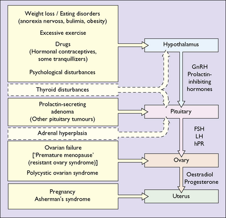
Table 28.1 Causes of secondary amenorrhea
Cause | Incidence (%) |
---|---|
Weight loss, low body weight, exercise | 20–40 |
Polycystic ovarian syndrome | 15–30 |
Pituitary insensitivity (post-pill) | 10–20 |
Hyperprolactinaemia | 10–20 |
Primary ovarian failure | 5–10 |
Asherman’s syndrome | 1–2 |
Hypothyroidism | 1–2 |
As noted in Chapter 1, normal menstruation depends on a normal uterus and vagina, and on the reciprocal interaction between hormones released from the hypothalamus (gonadotrophin-releasing hormones), the pituitary (the gonadotrophins – follicle-stimulating hormone (FSH) and luteinizing hormone (LH)) and the ovaries (oestrogen and progesterone).
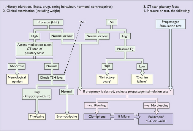
Investigation of secondary amenorrhoea
Unless organic disease is suspected, or the woman is desperately seeking relief from infertility, most experts would not investigate amenorrhoea until it has lasted for 6–12 months, as most women start menstruating during this time.
When investigation is indicated, a careful history is essential in which the doctor inquires about the woman’s general health, and seeks to determine whether she has an eating disorder or exercises excessively, or if any medical or psychiatric condition is present. A physical examination, including a vaginal examination, follows and in certain cases a pelvic ultrasound examination may be performed. If these examinations reveal no definite diagnosis, the following tests are ordered:
The purpose of the investigations is to exclude organic disease (for example a prolactin-secreting microadenoma or hypothyroidism) and to treat anovulation as a cause of infertility. If organic disease is not detected and infertility is not a problem, amenorrhoea does not represent a danger to the woman, but because low oestrogen levels may lead to bone loss, after 6 months of amenorrhoea hormone replacement treatment (see p. 325) should be advised.
More common causes of amenorrhoea
Weight loss
Women who have an eating disorder, particularly anorexia nervosa, cease to menstruate, as do some women who are compulsive exercisers. Amenorrhoea can occur in obese and overweight women who lose weight rapidly. The cause is a failure of the hypothalamus to release sufficient gonadotrophin-releasing hormone (GnRH) to initiate the release of gonadotrophins, and in consequence only a small quantity of oestrogen is secreted by the ovaries. If this persists for more than 6 months, bone loss may occur at a time when bone formation is reaching its peak. A consequence of this is a greater risk of osteoporosis in later life. Even during the recovery phase menstruation may not occur for several months, which may aggravate the problem.
Hyperprolactinaemia and prolactin-secreting tumours
Prolactin secretion by the pituitary gland is inhibited under normal conditions by dopamine released from the hypothalamus. In certain circumstances, this control is diminished. Examples are hypothyroidism and the administration of dopamine-depleting agents or dopamine receptor-blocking agents. A more common cause of hyperprolactinaemia is a microadenoma of the pituitary gland. In these cases, the increased circulating levels of prolactin act directly on the hypothalamus to reduce the secretion of GnRH, which in turn prevents the FSH and LH rises needed for follicle development and ovulation.
The woman develops oestrogen deficiency, with menstrual disturbances (usually amenorrhoea), a dry vagina and, often, a reduction of her libido. If the hyperprolactinaemia persists, osteopenia and, perhaps, osteoporosis will result. In 30% of women inappropriate milk secretion (galactorrhoea) occurs.
As hyperprolactinaemia accounts for 10–20% of cases of amenorrhoea, investigation is important. The diagnosis is made if a raised blood level of prolactin is found. If this is noted during investigation, a high-resolution CT scan of the pituitary is made to detect a prolactin-secreting tumour, although in most cases none is found. A microadenoma (<10 mm in diameter) is more common than a macroadenoma.

Stay updated, free articles. Join our Telegram channel

Full access? Get Clinical Tree

