Dental Emergency Care
Howard L. Needleman and Brian Grove
OROFACIAL TRAUMA
 EPIDEMIOLOGY
Trauma to children’s teeth is a very common event.1 The prevalence of these injuries varies depending on the population studied and the types of injuries reported. Studies indicate that as many as 46% of children sustained traumatic injuries to their primary or permanent teeth during childhood.2-4 Approximately 2% of children sustain such injuries annually.5 The majority of the injuries occur to the maxillary incisors due to their prominence in the dentition. Displacement injuries are more common in the primary dentition, because supporting bone in younger children is more flexible and pliable, while fractures are more common in the permanent dentition.
 RISK FACTORS
Several factors influence the child’s individual risk of sustaining traumatic injuries to the orofacial complex. Males are more likely to sustain injuries,6 while the frequency and type of injuries varies with age.7 The greatest incidence of trauma to the primary dentition occurs at 2 to 3 years of age, when motor coordination is developing.8 The incidence peaks again between the ages of 8 and 10 years, which poses a risk to the maxillary anterior teeth.8 Children who are very active, such as those with attention deficit hyperactivity disorder (ADHD)6,9or those with poor motor coordination such as with cerebral palsy,10 have greater risk of trauma. Socioeconomic status can also affect the risk of trauma to the dentition.6,11 Being a member of a non-nuclear family (ie, without two parents) will increase the risk of trauma.12 Children who are overweight12 and those with protrusive maxillary incisors13 are at greater risk of trauma. Individuals who have undergone general anesthesia with endotracheal intubation can experience “silent trauma” to their incisors (fractured or traumatized incisors during intubation).14 Failure to treat fractured teeth can impact a child’s daily performance, specifically in smiling, laughing, and showing teeth without embarrassment.15
 CLINICAL MANAGEMENT
The most common injuries to permanent teeth occur secondary to falls, violence, traffic accidents, and sports.4 Most sporting activities have an associated risk of orofacial injuries due to falls, collisions, and contacts with hard surfaces or other players. There are numerous preventive measures to decrease these risks, such as wearing protective intraoral mouth-guards and helmets during many activities. Two types of mouthguards are recommended, depending on the stage of the child’s dentition. The “boil and bite” mouthguard (eFig. 375.1  ) is inexpensive and can be adapted to the child’s mouth by the parent. These types of mouthguards are especially helpful during the mixed dentition, when primary teeth are exfoliating and permanent teeth are erupting, as they require frequent replacement. The custom-made mouthguard (eFig. 375.2
  ) is preferred due to its excellent fit but is more expensive, since it requires dental impressions and laboratory fees for fabrication. Early orthodontic intervention to reduce a severe protrusion of the maxillary incisors also has shown to decrease trauma to these teeth.17
In all instances of trauma and infection in an otherwise stable patient, a careful medical history, including tetanus vaccination, should be obtained.18 The skull, facial bones, and mandible should be palpated to assess any areas of ecchymosis, paresthesia, crepitus, or pain, which may point to a concomitant facial bone fracture. In addition, a subsequent cranial nerve function test should be performed. Finally, to rule out aspiration in cases of a tooth fracture or avulsion, where the tooth or fragment cannot be located, immediate chest and abdominal radiographs should be obtained.18
Orofacial trauma, including trauma to the dentition, is common in cases of child abuse and neglect.19-22 Dental trauma may be an important marker for child abuse, because craniofacial, head, face, and neck injuries occur in approximately 65% of recorded cases of child abuse.21 Injuries that have typical shapes and patterns, human bite marks, adult handprints, or bilateral injuries indicate possible abuse.
Typical oral injuries include lacerations, especially of the maxillary labial frenum (Fig. 375-1); fractured, luxated, or avulsed teeth; jaw and facial fractures; oral burns; oral and facial bruises; and tissue scarring from previous trauma. Teeth may be discolored or abscessed from previous trauma. Sexually transmitted oral lesions such as condyloma acuminata (eFig. 375.3  ) in prepubertal children should raise suspicion of sexual abuse. Oral trauma from sexual abuse can also result in petechiae or bruising at the junction of the hard and soft palate (Fig. 375-2). Burns and bruising on the lips may also be present and could be the possible sequelae of forced feeding.24
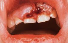
FIGURE 375-1. Tear of the maxillary labial frenum.
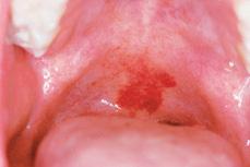
FIGURE 375-2. Petechiae of the palate secondary to sexual abuse.
Soft Tissue Trauma
Trauma to the soft tissues of the oral cavity can be caused by physical, chemical, or electrical insults. Soft tissue injuries consist of abrasions, lacerations, contusions, ecchymoses, hematomas, and burns. Oral lacerations should be examined carefully for the presence of foreign bodies, especially in the presence of fractured teeth (eFig. 375.4A,B  ). A radiograph of the lesion should be done to rule out foreign bodies, as visual inspection and palpation alone are usually not sufficient (eFig. 375.5A,B
  ). Lip lacerations require careful management to provide an esthetic closure, especially if the laceration is deep or extends through the vermilion border. Full-thickness lacerations require suturing in layers. Careful attention to anatomic alignment of the vermilion border is important. Through and through lacerations result in communication between the skin and oral environment and are frequently contaminated. Suturing of the intraoral laceration should precede skin suturing, and the patient should be placed on a course of antibiotics effective against staphylococcal organisms. Tongue lacerations are commonly seen in children and usually result from a fall or blow to the chin. The tongue has a profuse blood supply, and injury can result in copious bleeding. Most tongue lacerations with approximating borders will heal without suturing; however, tears that leave unapproximated borders, such as at the tip or along the lateral borders, require suturing. Gingival degloving occurs when both the gingival tissue and periosteum are pulled away from its normal position around the tooth, exposing underlying bone (Fig. 375-3). These injuries require careful repositioning of the gingival tissue and stabilization with sutures.
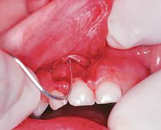
FIGURE 375-3. Degloving of the maxillary labial gingival.
Electrical Burns Electrical burns of the mouth are an infrequent but serious event with varied interdisciplinary treatment approaches.25 These injuries, which affect children mostly under the age of 3, typically occur when a child sucks on the end of an extension cord or bites through the insulation of a live wire. The best way to avoid these accidents is to take preventive measures against them by using safety caps on all electrical outlets and heavy, solid insulation on all electrical cords. Immediate treatment should address systemic complications such as shock and hemorrhage. The wound requires careful daily débridement of the necrotic tissue, approximation of the wound edges with adhesive straps, and topical antibiotics. Complications may result from electrical burns of the oral commissure such as bacterial infection, disfiguration, microstomia, and discoloration of teeth.27 Labial artery bleeding is a late complication of oral commissure burns.27 If the oral commissure is involved, using an intraoral splint (eFig. 375.6A,B  )28to prevent labial adhesions and to limit the oral opening can decrease the need for commissuroplasty.29
Tooth Injuries
Injuries to teeth can be divided into fractures and displacements. The International Association of Dental Traumatology has established guidelines for their evaluation and management.30 Fractures of teeth are classified by the extent of the lost tooth structure. Most of these injuries require a thorough extra- and intraoral examination as well as dental radiographs. Both intraoral and extraoral radiographic evaluation is important to fully evaluate the extent of the injury. In addition, a baseline radiograph is essential for comparison to later radiographs to evaluate healing and the status of the periapical tissues, should symptoms of pulpal necrosis arise.
Stay updated, free articles. Join our Telegram channel

Full access? Get Clinical Tree


