Class Ia
Investigations capable of accurately identifying congenital uterine anomalies and classifying them into appropriate subtypes (accuracy >90 %):
Hysteroscopy and laparoscopy
SHG
3D US
Class Ib
Investigations capable of accurately identifying congenital uterine anomalies without being able to classify them into appropriate subtypes (accuracy >90 %):
Hysteroscopy alone
Class II
Investigations capable of identifying congenital uterine anomalies (accuracy <90 %):
HSG
2D US
Class III
Investigations of which the accuracy in diagnosing congenital uterine anomalies is uncertain:
MRI
Physical examination during pregnancy or delivery
Traditionally, diagnostic laparoscopy is considered the best complementary examination to hysteroscopy, and the combination hysteroscopy/laparoscopy is accepted as the “gold standard” in evaluating congenital uterine anomalies. Hysteroscopy with laparoscopy offers the added advantage of concurrent treatment, as in the case of a uterine septum resection.
However, laparoscopy is an invasive and expensive one, therefore the scientific efforts have recently focused at allowing the differential diagnosis of the various anomaly subtypes without it. Furthermore, with the new ESHRE/ESGE classification and the need to measure fundal, septal and lateral uterine wall thicknesses, it could be hypothesized that the actual gold standard test may be replaced by another imaging modality in the future. Indeed the laparoscopic approach does not always allow accurate and objective uterine measurements. Furthermore, another theoretical limit of any endoscopic imaging technique, including either hysteroscopy alone or the current hysteroscopic and laparoscopic “gold standard” approach, is that they are based only on the subjectively impression of the clinician who performs the examination and do not always allow accurate and objective uterine measurements.
Various types of ultrasound examinations are nowadays available for the diagnostic of female genital anomalies [18, 19]. They have the advantages of being non-invasive, easily accessible and well-accepted form the patients. With the use of various ultrasound techniques measurable and objective estimations of uterine wall, uterine cavity and the external uterine contour could be done.
Two dimensional ultrasound is simple, available in almost every outpatient clinic, and can give reliable, reproducible and measurable informations on uterine anatomy, leading to the exact diagnosis as well as the differential diagnosis between the different categories (See Chap. 6). However, it has a lower diagnostic accuracy in comparison with the other sonographic techniques (Table 10.1) [18–20].
Hystero-contrast-sonography (HyCoSy) by using the contrast medium in the uterine cavity offers the additional advantage of a better internal delineation of the uterine contour, providing additional information of the morphology of the uterine cavity; furthermore, it is an office procedure, with low risk and high patient satisfaction rates (See Chap. 6) [21]. However, some patients experience some degree of pain, which is however reduced compared to HSG or hysteroscopy. The diagnostic accuracy of this method is estimated to be higher, with a sensitivity and specificity of 93 and 99 % respectively [18] (Table 10.1).
Three dimensional Ultrasound is a non invasive and highly reproducible method of investigation, which provides the simultaneous view of the three planes of the uterus, along with the complete volume scan (See Chap. 7) [18, 22]. Due to its highest diagnostic accuracy (Table 10.1) it seems that it could be the new “gold standard” in the diagnosis of female genital malformations especially the uterine ones.
Recently, new data are emerging regarding the integration of hysteroscopic findings with 3D US data. This trend is motivated by safety and non-invasiveness of 3D US and by its high accuracy (class IA). Indeed, 3D US in expert hands enables clear visualization of the uterine fundus and investigation of coexisting adnexal disease [18, 22].
Magnetic resonance imaging (MRI) (See Chap. 8) is a relatively sensitive tool (Table 10.1) and some authors suggest that it could supply invasive procedures such as hysteroscopy and laparoscopy for the diagnosis of a malformed uterus, especially in cases of adolescent patients and/or children in which the diagnosis is performed for other reasons than infertility (ie pelvic pain, menstrual abnormalities etc). The disadvantages of this technique are that it is expensive and not available in many clinical contests [18, 19].
The evidence to date suggests that several investigations have a satisfactory overall accuracy in diagnosing the presence of a female genital tract congenital anomaly. The most accurate investigations in order seems to be: (i) 3D US, (ii) HyCoSy, (iii) MRI, (iv) 2D US and (v) HSG [8].
However, it seems that conclusions as to which investigations are able to correctly sub-classify the anomalies could not be considered as completely final. This is due to the fact that, prior to the ESHRE/ESGE classification, in the absence of clear definitions, there was no unanimous agreement how to objectively distinguish between the normal and the arcuate uterus, the arcuate and the septate uterus, the bicornuate and the didelphys uterus and the combined septate bicornuate uterus.
As the concordance between 3D US and combined hysteroscopy and laparoscopy appears to be the high (Table 10.1), and considering the need to obtain accurate measurements of the uterine walls in order to make the appropriate diagnosis (as highlighted in the new ESHRE/ESGE classification), it appears that the new gold standard method of choice should be the 3D US.
A Diagnostic Algorithm for Female Genital Tract Anomalies: Proposals
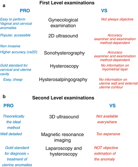
Looking for a diagnostic algorithm for female genital tract anomalies, it is out of doubt that all the available first- and second- line diagnostic tools used for diagnosing such anomalies present advantages and disadvantages and that, generally, the final diagnosis is possible only by integrating two or more examinations (Fig. 10.1).