Fig. 49.1
Chest x-ray of a child with a paraesophageal hernia showing herniation of bowel loops in addition to the stomach (a) and a lateral chest x-ray showing bowel herniation in a large paraesophageal hernia (b)
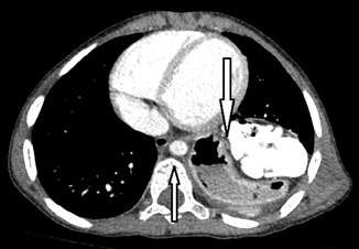
Fig. 49.2
CT scan showing a large paraesophageal hernia containing stomach and bowel loops
Etiology
The exact etiology of congenital paraesophageal hernia is not known .
It is postulated that congenital paraesophageal hernia is secondary to embryonal developmental defects in the lumbar component of the diaphragm leading to defective right crus of the diaphragm.
Most reported cases of congenital paraesophageal hernia occur sporadically.
A familial occurrence of hiatal hernia was first suggested in 1939.
Since then, there have been several reports documenting the occurrence of hiatal hernia among siblings. This unusual familial occurrence supports a genetic predisposition to the development of congenital paraesophageal hernia and an autosomal dominant mode of inheritance was suggested .
Classification
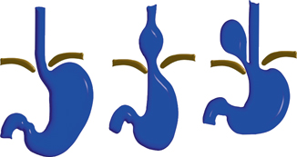
Fig. 49.3
Diagrammatic representation of the different types of hiatal hernias
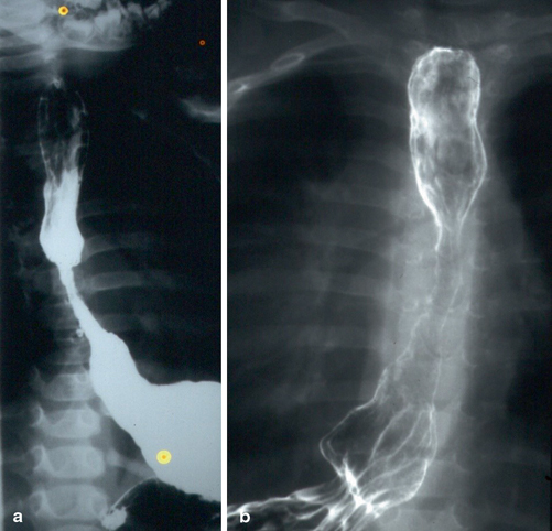
Fig. 49.4
a and b Barium swallow showing sliding hiatal hernia with proximal stricture formation. Note also part of the stomach above the gastroesophageal junction
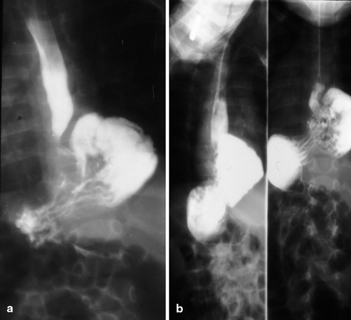
Fig. 49.5
a and b Barium meal showing paraesophageal hernia
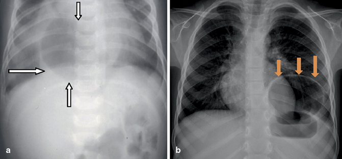
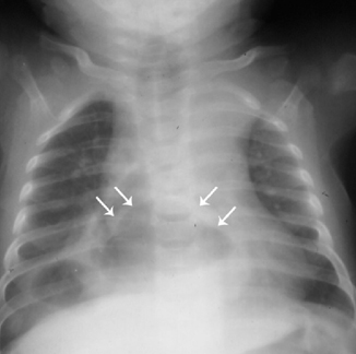
Fig. 49.6
Chest x-ray showing paraesophageal hernia in a child. a, Note the gas in the herniated part of the stomach into the chest. b, Note the hernia sac containing stomach