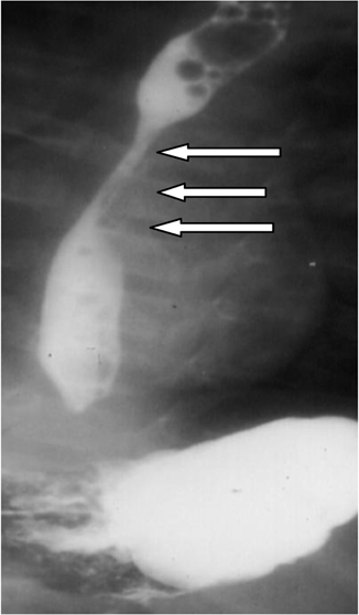
Fig. 16.1
Congenital esophageal stenosis (left arrows) in a postoperative patient with esophageal atresia and tracheoesophageal fistula (right arrows)

Fig. 16.2
Congenital esophageal stenosis affecting the middle third of the esophagus (arrows)
It is important to rule out other diagnosis like achalasia and stricture secondary to gastroesophageal reflux
Esophageal pH monitoring is valuable in this regard.
Investigations such as pH monitoring, manometry, and endoscopic biopsy are performed to rule out other conditions mimicking CES such as acquired esophageal stenosis and achalasia. It is not uncommon for achalasia to be confused with CES.
Radiologically, it is difficult to differentiate between the three types but membranous (web) obstruction is more localized affecting a much shorter segment, but in the presence of severe proximal dilatation this may be difficult to demonstrate.
Stay updated, free articles. Join our Telegram channel

Full access? Get Clinical Tree


