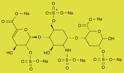
Fig. 17.1
William Halsted, he performed truncal blocks with cocaine injections, but he died as cocaine addicted

Fig. 17.2
James Leonard Corning (1855–1923), he was an American neurologist, mainly known for his early experiments on neuraxial blockade

Fig. 17.3
Gaston Labat. Needles for spinal anesthesia employed at the time of Labat
Regional anesthesia is extremely safe, but, like all of surgical procedures, it is not completely safe and carries risks. However, serious complications related to obstetric neuraxial anesthesia occur rarely but can be devastating when they occur. There have been various case reports, case series, or retrospective studies, but the lack of large comprehensive database makes difficult an accurate estimation of the incidence of complications. In recent decades the development of maternal anesthesia practice, either spinal or epidural block, has improved pregnancy safety, particularly in women undergoing cesarean delivery, living in rural areas, or having preexisting medical comorbidities [2, 3]. The purpose of this chapter is to discuss the complications occurring during regional anesthesia, their diagnosis, and treatment.
Before to start, please remember the correct and pathological anatomy of the column, before and during pregnancy (Figs. 17.4, 17.5, 17.6a, b, and 17.7).


Fig. 17.4
The correct and pathological anatomy of the column: the lumbar vertebrae

Fig. 17.5
The bones of the vertebral canal and its angle


Fig. 17.6
(a) Example of adult spine that presents pathological curvatures. (b) Example of adult spine that presents pathological curvatures

Fig. 17.7
The sacrum in anterior views and its angles
17.2 Cardiovascular Complications During Regional Anesthesia
17.2.1 Hypotension
Hypotension is almost inevitable adverse complication of spinal anesthesia induced by sympathetic block, which causes a significant decrease in the venous return and cardiac output due to systemic vascular resistance reduction and venous capacitance increase.
In the literature there is not one accepted definition of hypotension and the incidence of hypotension varies between 1.9% and 71%, depending on the chosen definition. The most frequently applied definition are systolic arterial blood pressure below 100 mmHg or a decrease below 80% from baseline (Fig. 17.8).


Fig. 17.8
Maternal hypotension (systolic arterial pressure of 100 mmHg or less in the left) after spinal anesthesia. The patient is positioned in lateral and safe position to reduce hypotension (in the right) (modified from: Antonio Malvasi Gian Carlo Di Renzo, Semeiotica Ostetrica. C.I.C. International Publisher, Rome, Italy, 2012)
The higher level of block (T4) (Fig. 17.9) required for cesarean delivery, the anatomic and physiologic changes (Fig. 17.10) of parturients, and the reduced sensitivity to the endogenous vasoconstrictors associated with increased synthesis of endothelium-derived vasodilators, determining an increased susceptibility to the effects of sympathectomy, expose the parturients to higher risk of hypotension (Fig. 17.11) [4, 5].


Fig. 17.9
The different level of block after injection of drug in subarachnoid space and different anesthetic drugs distribution in the spinal channel

Fig. 17.10
Evaluation of the anatomic and physiologic changes of column

Fig. 17.11
The physiologic changes of parturients post T4 spinal block
Nausea and vomiting (Fig. 17.12) are frequently associated with hemodynamic changes induced by spinal blockade. A decrease in blood pressure at the critical level, compounded by aortocaval compression, may compromise uteroplacental blood flow, leading to fetal hypoxia and acidosis. Risk factors for hypotension are maternal body mass index ≥29 kg/m2, age ≥35, hypertension, higher fetal weight, associated comorbidities, and local anesthetic injected. Therefore, in the literature local anesthetics dose has been widely studied in order to find out the right matching between hemodynamic changes and perioperative analgesia quality [6–15].


Fig. 17.12
Maternal nausea and vomiting are the most common symptoms after spinal block (modified from: Antonio Malvasi Gian Carlo Di Renzo, Semeiotica Ostetrica. C.I.C. International Publisher, Rome, Italy, 2012)
In same cases during cesarean section, the uterine exteriorization, uterine replacement in the abdominal cavity, and other maneuvers on the bowel produce the same nausea and vomiting (Fig. 17.13).


Fig. 17.13
Maternal nausea and vomiting occur during uterine exteriorization, uterine replacement in the abdominal cavity, and other maneuvers on the bowel (modified from: Antonio Malvasi Gian Carlo Di Renzo, Semeiotica Ostetrica. C.I.C. International Publisher, Rome, Italy, 2012)
Indicators for predicting alterations of autonomic function, leading to an increased risk of hypotension under spinal anesthesia, are positional blood pressure and heart rate changes between the left lateral to supine position [16]. However, bedside test can be associated to the promising advanced techniques as the assessment of heart rate variability performed on the same day of surgery, although it lacks standardization [14].
Maternal hypotension can be prevented by intravenous fluid preloading (Fig. 17.14) and left uterine displacement to avoid aortocaval compression (Figs. 17.15 and 17.16), associated with vasopressor therapy (Table 17.1) [17].


Fig. 17.14
Maternal bradycardia (a) and hypotension (b) can be prevented by intravenous fluid preloading (c)

Fig. 17.15
Lateral position of parturient after spinal anesthesia with wedge pillow to displace the uterus on the left (modified from: Antonio Malvasi Gian Carlo Di Renzo, Semeiotica Ostetrica. C.I.C. International Publisher, Rome, Italy, 2012)

Fig. 17.16
Left uterine displacement to avoid aortocaval compression and prevents maternal hypotension after spinal anesthesia
Table 17.1
Prevention and treatment of hypotension in parturients undergoing elective cesarean delivery with spinal anesthesia
Prevention and treatment of hypotension |
|---|
Fluid therapy |
Timing of administration: preload – coload |
Type of fluid: crystalloid, colloid |
Vasopressor |
Lower anesthetic doses |
Leg elevation or wrapping |
Elastic stockings |
Left uterine displacement |

Fig. 17.17
Phenylephrine and its most important effects

Fig. 17.18
Ephedrine and its most important effects, as cardiac output and blood pressure increasing

Fig. 17.19
Methoxamine and its most important effect, as augmentation of myocardial contractility and reduction of fetal oxygenation

Fig. 17.20
Mephentermine and its most important effects

Fig. 17.21
Metaraminol and its most important effects

Fig. 17.22
Autonomic cardiac innervation (parasympathetic and sympathetic branches) and the heart effects

Fig. 17.23
Example of fetal bradycardia

Fig. 17.24
Maternal complications: valvular heart disease and cardiac arrest after hypotension – spinal block related

Fig. 17.25
Post-dural puncture headache and other complications

Fig. 17.26
Acetaminophen

Fig. 17.27
Caffeine and sumatriptan, a serotonin type 1-d receptor agonist, have cerebral vasoconstriction effects

Fig. 17.28
Gabapentin

Fig. 17.29
Pregabalin

Fig. 17.30
Epidural abscess and microbiological culture

Fig. 17.31
The importance of using and documenting meticulous aseptic technique

Fig. 17.32
The use of masks, surgical cap, sterile gloves, a sterile gown, and aseptic technique is required for a sterile environment

Fig. 17.33
The presence of tattoo as a contraindicated place for insertion of catheter

Fig. 17.34
(a–l) Epidural catheter placement: under aseptic conditions (a–c), epidural catheter placement was performed in the sitting position using the midline approach: tissue infiltration to the ligament flavum was performed using 5 ml of 1 % lidocaine (d). After, in the sitting position, a 20 G epidural catheter was inserted using an 18-gage Tuohy needle at the L3–L4 level interspace. The epidural space was identified using the loss of resistance technique with saline solution (e–h). After negative aspiration for cerebrospinal fluid and blood, a 3 ml test dose of 2 % lidocaine with adrenaline was given over 15 s through the catheter. Before insertion, the catheter and bacterial filter were primed with physiologic saline (i–l), aiming to achieve an air-free column of fluid from the connector to the catheter tip. If no sign of an intravascular or subarachnoid puncture was observed, the catheter was secured and the parturient was placed in the supine position, and after the main dose of fentanyl (100γ) and levobupivacaine (0.0625 %) diluted with isotonic sodium chloride solution to a volume of 15–20 ml was injected. When labor protracted, additional doses of drugs were administered at hourly intervals on the indication of maternal request. Two hours after childbirth, the epidural catheter was removed

Fig. 17.35
Significant epidural hematoma

Fig. 17.36
Heparin

Fig. 17.37
Warfarin

Fig. 17.38
Aspirin

Fig. 17.39
The thromboelastography

Fig. 17.40
(a) Waveform generated by TEG analyzer. (b) Normal value of thromboelastography in healthy obstetric patient

Fig. 17.41
Normally the conus medullaris ends near lumbar vertebral levels 1 (L1), occasionally lower. At the level of L1, the spinal nerves continue to branch out diagonally, forming the cauda equina

Fig. 17.42
Lidocaine

Fig. 17.43
Epidural catheter erroneously inserted in the subarachnoid space (modified from: Antonio Malvasi Gian Carlo Di Renzo, Semeiotica Ostetrica. C.I.C. International Publisher, Rome, Italy, 2012)

Fig. 17.44
Progression of local anesthetic systemic toxicity

Fig. 17.45
Propofol: chemical formula (a) and commercial presentation (b)

Fig. 17.46
Methylene blue

Fig. 17.47
Transabdominal transversal sonographic scan of the fetal head in left occiput posterior position during the second labor stage in epidural analgesia. The picture shows the fetal orbits, the lens, the nasal bridge, and the occipital bone in the left pelvis
One of the foremost methodologies is the prophylactic administration of 10–20 ml/kg intravenous fluids over 15 min. Colloid solution would be more effective as compared to crystalloids due to their higher colloid osmotic effect and a longer half-life in the intravascular space, but preloading of colloids is not popular routinely due to risk of anaphylactoid reactions, derangement of coagulation, suppression of platelet activity, and increased costs. On the other hand, large volume of crystalloids may induce the atrial natriuretic peptide secretion contributing to peripheral vasodilatation [18]. According to results from the general population receiving spinal anesthesia and due to studies establishing that maternal hypotension occurs in the period just following the spinal injection, the rapid coloading administration of fluid (15 ml/kg) initiated immediately on induction of spinal anesthesia had also gained a widespread acceptance in obstetric fluid management, especially during emergent conditions [18–19]. However, a slower speed of injection and lower dose of spinal anesthetic resulted in a lower incidence of hypotension or delayed onset [14].
Intravenous vasopressors can be used to treat hypotension in laboring women without adverse effect on the fetus (Table 17.2) [20]. In the absence of maternal bradycardia, phenylephrine may be preferable because it increases systemic vascular resistance more than ephedrine. Conversely, ephedrine raises stroke volume and heart rate more than phenylephrine but impairs fetal acid-base status stimulating fetal beta-adrenergic receptors with consequent fetal tachycardia and increased metabolic demand [21].
Table 17.2
Comparative analysis of vasopressors used in obstetric anesthesia
Drug | Mechanism of action | Advantage | Disadvantage |
|---|---|---|---|
Phenylephrine (Fig. 17.17) | Selective α1 receptor agonist | Peripheral vasoconstriction, raised systemic vascular resistance, and arterial blood pressure Immediate onset and short duration of action Ideal for continuous infusion No adverse effect on fetal acid-base status as compared to ephedrine | Tachyphylaxis Reflex bradycardia can decrease maternal cardiac output |
Ephedrine (Fig. 17.18) | α, β receptor agonist. Both direct and indirect effect on the sympathetic nervous system | Increased myocardial contractility (β1 receptor) Peripheral arterial and venous vasoconstriction (α receptor) Release endogenous norepinephrine (indirect effect) Does not need multiple dilutions as compared to phenylephrine | Tachyphylaxis Adverse effect on fetal Acid-base status |
Methoxamine (Fig. 17.19) | α1 receptor agonist | Peripheral vasoconstriction No inotropic or chronotropic effect Rare cases of tachyphylaxis | Reflex bradycardia Adverse effect on fetal acid-base status |
Mephentermine (Fig. 17.20) | α receptor agonist. Both direct and indirect effect | Peripheral vasoconstriction (α receptor) Release endogenous norepinephrine (indirect effect) Does not need multiple dilutions as compared to phenylephrine | Tachyphylaxis |
Metaraminol (Fig. 17.21) | α receptor agonist. Both direct and indirect effect | Peripheral vasoconstriction (α receptor) Release endogenous norepinephrine (indirect effect) No adverse effect on fetal acid-base status as compared to ephedrine | Tachyphylaxis |
Other authors did not observe the difference between the two vasopressors in the incidence of fetal acidosis and confirmed the higher incidence of bradycardia, due to baroreceptor reflex response, in patients receiving phenylephrine which is a selective α1-adrenergic receptor agonist [22]. Since the optimal dosing regimen is unclear, novel closed-loop double pump automated systems are developed to automatically administer vasopressors, based on continuous noninvasive arterial pressure monitoring with remarkable benefits on maternal hemodynamic stability during cesarean delivery [23]. Noninvasive cardiac output monitoring might be indicated for patients with severe cardiac disease, while the decision to perform invasive hemodynamic monitoring should be based on individual patient’s medical history and cardiovascular risk factors [14, 19].
Rarely, severe hypotension with bradycardia and cardiovascular collapse can occur after central neuraxial anesthesia, secondary to activation of the cardioinhibitory reflex called Bezold-Jarisch reflex. Prompt treatment with adequate doses of vasopressors is required. If cardiac arrest does occur, cardiopulmonary resuscitation should immediately start, and the fetus should be rapidly delivered in order to improve both maternal by alleviating aortocaval compression and fetal survival prospects. At delivery oxytocin should be administered as uterotonic but must be cautiously administered because of its systemic vasodilatation and coronary vasoconstriction [24].
17.2.2 Bradycardia and Cardiac Arrest
Bradycardia and cardiac arrest are the most feared complications, whose incidence is higher with spinal anesthesia than general anesthesia. The blockade of the preganglionic cardioaccelerator fibers originating between T1 to T4 may progress to complete heart block or asystole (Figs. 17.22, 17.23, and 17.24). It is also aggravated by decreased preload after spinal anesthesia leading to decreased action of right atrial stretch receptors.
Intravenous atropine is typically used as the first line of therapy and also for prophylaxis; ephedrine is used when hypotension is associated with bradycardia or in unresponsive cases to atropine. Cardiovascular side effects are often treated when the parameters vary more than 20 % from their baseline, if the systolic blood pressure is less than 100 mmHg or if the patient becomes symptomatic (e.g., nausea or faintness) [25].
17.3 Post-dural Puncture Headache
Post-dural puncture headache (PDPH) is troublesome iatrogenic complication of unintended dural puncture during epidural or spinal technique, mostly observed in parturients population with an incidence between 0 and 2.5 % [25]. The depletion of cerebrospinal fluid (CSF) volume, by leaking through the dural hole, causes a decrease in the intracranial pressure, stretching of pain-sensitive cranial structures, and may induce a compensatory cerebral vasodilatation according to the Monro-Kellie doctrine [26]. The experience of the anesthesiologist, patient position, and type and size of needle do not appear to influence the incidence of PDPH as recently observed [27]. PDPH is usually severe, positional, and localized in frontotemporal-occipital regions, radiating to the neck and shoulder. Nausea, vomiting, vertigo, tinnitus and hyperacusis, photophobia, and diplopia (due to traction on the sixth cranial nerve) might be associated with headache (Fig. 17.25). Two cases of thoracic back pain without headache have been described [28].
Stay updated, free articles. Join our Telegram channel

Full access? Get Clinical Tree


