Fig. 41.1
Type I (a), type II (b) and type III (c) choledochal cyst
IA: Fusiform dilatation of the entire extrahepatic bile duct
IB: Fusiform dilatation of a segment of the extrahepatic bile duct
IC: Fusiform dilatation of the CBD portion of the extrahepatic bile duct
Type II: isolated diverticulum protruding from the CBD. This comprises >5 % of all types of choledochal cysts (Fig. 41.1b).
Type III or choledochocele: cystic dilatation of the intraduodenal portion of the extrahepatic CBD (choledochocele). It comprises approximately 5 % of all choledochal cysts (Fig. 41.1c).
Type IVa: characterized by multiple dilatations of the intrahepatic and extrahepatic biliary tree (Fig. 41.2).
Fig. 41.2
Type IVa (a) and V (b) choledochal cyst
Type IVb: Multiple dilatations involving only the extrahepatic bile ducts.
Type V or Caroli’s disease: cystic dilatation of intra hepatic biliary ducts (Fig. 41.3).
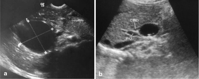
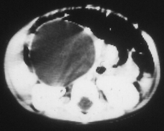
Fig. 41.3
Abdominal ultrasound showing choledochal cyst. Note also the portal vein and proximal part of the common bile duct
The term forme fruste choledochal cyst was proposed by Lily et al. to describe one of these variants in which the cystic dilatation of the common duct is minimal or absent but there is a long pancreatobiliary union, partial obstruction of the lower common bile duct, and histopathological changes identical to choledochal cyst in the wall.
Clinical Features
The classic triad for choledochal cysts is:
Pain
Jaundice
Abdominal mass
This is found in only a minority (20–60 %) of children at the time of presentation.
Infants commonly present with:
Elevated conjugated bilirubin (80 %)
Failure to thrive
An abdominal mass (30 %)
In patients older than 2 years of age:
Abdominal pain is the most common presenting symptom
This is usually associated with intermittent jaundice
Recurrent cholangitis and pancreatitis
The most common complications of a choledochal cyst are:
Cholangitis, pancreatitis, biliary cirrhosis, liver abscess, cholelithiasis, portal hypertension, cyst rupture, malignant degeneration.
The risk of complications increases with age and the most important complication is malignant degeneration, with an incidence of 2.5–26 %.
Diagnosis