Cat Scratch Disease (Bartonella henselae)
Moshe Ephros and Michael Giladi
Cat scratch disease (CSD) is a ubiquitous, self-limited infection characterized by prolonged regional lymphadenitis and often an inoculation site papule, usually after a cat’s scratch or bite, and caused primarily by Bartonella henselae. In 10% to 20% of cases, the lymph node will suppurate.1,2 In a minority of cases (approximately 10%), a wide range of extranodal manifestations collectively known as atypical CSD may occur, including fever of unknown origin, as well as visceral, neurologic, and ocular involvement.
 EPIDEMIOLOGY AND PATHOPHYSIOLOGY
CSD occurs worldwide and is prevalent in warm and humid climates. In temperate zones, CSD occurs primarily during fall and winter, sometimes also during the summer, while in the tropics, it occurs throughout the year.1-3 In 2000, the US annual hospitalization rate was estimated at 0.6/100,000 for children under 18 years old; not surprisingly 24% of admissions were for atypical disease.9 Cats, especially kittens, are the major reservoir and tend to have prolonged, asymptomatic bacteremia. They infect humans by scratch, bite, or mucous membrane inoculation.2,5,10-14 The cat flea is associated with increased cat infectivity although it probably does not play a major role in cat-to-human B henselae transmission.5,11,15 Direct human-to-human spread has not been reported; however, indirect transmission of B henselae by red cell transfusion may be possible.19
Bartonellae are fastidious, slow-growing, pleomorphic gram-negative bacteria. Culture of B henselae may require several weeks of incubation before colonies can be detected. Routine cultures are not likely to detect growth.  The pathogenesis of CSD is poorly understood, and the reasons some patients develop typical CSD with localized infection and regional necrotizing granulomatous adenitis while others develop serious atypical disease, presumably due to blood-borne dissemination with visceral or other end-organ involvement, is not clear. Bacterial and host factors play a role. The type and severity of the host immune response to infection also modify the clinical picture.26,27 Immunocompromised individuals, especially those with cell-mediated immune deficiency, either congenital or acquired (especially HIV-AIDS), are at risk of developing severe or atypical disease.2,20,28B henselae can infect and persist in CD34+ differentiating hematopoietic progenitor cells29; like other Bartonellae, it interacts with endothelial cells and is capable of inducing angiogenesis.30
 CLINICAL FEATURES
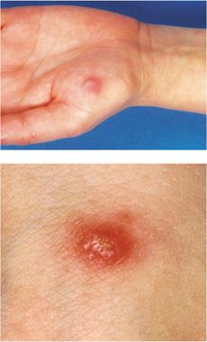
Cat scratch disease (CSD) is probably the most common cause of prolonged subacute regional lymphadenitis.2,33 Cutaneous inoculation of B henselae is 90% cat, especially kitten-associated, 50% to 75% of which is via scratch or bite.1,2,16,33 A papule often appears at the inoculation site and persists for a few weeks (Fig. 257-1).1,2,16,33 Most frequently, CSD involves a lymph node or a group of nodes draining the inoculation site.1,2 Usually tender initially, these nodes appear from 1 week to 1 to 2 months after inoculation and erythema may occur. Suppuration occurs in 10% to 20%.1,2,16,33 CSD is primarily an infection of the nodes draining the upper extremity, head, neck, and groin, but any lymph node location is possible (eFigs. 257.1 and 257.2  ).1,2,16,33 Between 30% and 50% of patients will have an elevated temperature that can be accompanied by malaise, anorexia, night sweats and headache.1,2,16 Resolution is usually over weeks to a few months, though rarely, recovery may be prolonged.
Typical CSD
 Maintaining a high index of suspicion and taking a detailed history, particularly that of cat contact, are important. Infection is most commonly the cause of regional adenopathy/adenitis: bacterial—for example, staphylococcal, streptococcal; mycobacterial for example, tuberculosis and especially atypical mycobacteria; fungal; viral for example, EBV, CMV, HIV; parasitic for example, toxoplasmosis. The possibility of malignancy, especially lymphoma, or of a benign tumor must be entertained. Noninfectious, nonneoplastic causes include sarcoidosis, Kawasaki disease, sinus histiocytosis with massive lymphadenopathy, congenital neck anomalies, autoimmune lymphoproliferative syndrome, histiocytic necrotizing lymphadenitis (Kikuchi disease), sometimes possibly due to B henselae,36 idiopathic facial aseptic granuloma, and drug-induced lymphadenopathy (eg, due to phenytoin).
Complicated CSD
In immune competent children, atypical (extranodal or complicated) CSD occurs in 10% to 15%.1,2,16,33 Occasionally, B henselae disseminates to liver, spleen, eye, CNS, bone, and other sites. The most common atypical presentation of CSD is Parinaud oculoglandular syndrome granulomatous conjunctivitis and preauricular lymphadenitis.2,37,38 Dissemination to viscera usually involves liver or spleen, often with prolonged fever and abdominal pain.2,39,40 Ultrasound and CT suggest microabscesses, which histologically are necrotizing granulomata. CSD is a common cause of FUO in children (eFig. 257.3  ).2,41-48 Eye manifestations include neuroretinitis, where the patient complains of acute, usually unilateral, visual deterioration; stellate macular exudates (“macular star”) are seen funduscopically. This is associated with sequelae in some, though most recover.2,37,42,43 Neurologic complications include encephalopathy, seizures and cranial nerve palsies.2,44-47 Lumbar puncture is either normal or with a mild, predominantly mononuclear pleocytosis, or mildly elevated CSF protein. CT is normal, and electroencephalogram (EEG) is encephalopathic. Other atypical manifestations include osteomyelitis, rashes (eg, erythema nodosum) and pneumonitis.59,60 Two very important kinds of B henselae infection not within the scope of this chapter are bacterial endocarditis,2,20,33,61 and infection of immunocompromised patients including vascular proliferative cutaneous and visceral lesions of bacillary angiomatosis and bacillary peliosis, and persisting or relapsing bacteremia.2,20,33
 DIAGNOSTIC EVALUATION
Historically, diagnosis was made when a patient presented with a cat scratch or bite, a primary inoculation lesion, regional lymphadenitis and a characteristic biopsy, sterile pus or a positive skin test with cat scratch antigen. Clinical suspicion in the appropriate epidemiological context is still extremely important, but given the expanded spectrum of CSD, specific diagnostic tools are required, especially when a classical history is absent. Routine laboratory tests are usually not helpful: WBC, ESR, CRP may be normal or elevated. Negative cultures of B henselae from blood or tissue are the rule rather than the exception. The most practical way to diagnose CSD is serologically. The IgG-based immunofluorescent antibody test (IFAT), such as that performed at the Centers for Disease Control and Prevention, has a sensitivity of 84% to 95% and a specificity of 94% to 98%.20 Results from studies performed in Europe are less satisfactory and inconsistent, perhaps due to higher background seroprevalence.62,63 Cross-reactivity with other organisms including Bartonellae (eg, B quintana) has been reported.20 Enzyme immunoassay (EIA) tests are also used, but variable sensitivity is also a problem.64 An outer-membrane protein-based EIA has a sensitivity of 85% (when IgG and/or IgM anti-B henselae antibodies are positive) and a specificity of 98% and can be useful in differentiating old from acute disease.65,66 Both IFAT and EIA antibodies develop slowly; thus, seroconversion may occur only after ≥3 weeks. Polymerase chain reaction (PCR) of lymph node or pus aspirate, primary skin lesions or of other tissue is a highly sensitive and specific diagnostic tool. Both broad-range and species-specific PCR assays have been developed. It is particularly useful when serology is negative and definitive diagnosis is urgent (eg, in those who are particularly ill, who have atypical disease or complications, whose lesions are not resolving, the immune compromised, or when other diagnoses such as malignancy are being entertained).67,68 Fine-needle aspiration of a lesion for PCR and cultures may suffice, although excisional biopsy is indicated when malignancy needs to be ruled out. One difficulty is in considering the diagnosis of CSD, particularly atypical CSD, in the absence of cat exposure or lymphadenopathy. 
Stay updated, free articles. Join our Telegram channel

Full access? Get Clinical Tree


