Fig. 42.1
Classification of biliary atresia
Type I: Atresia involving the common bile duct. The proximal bile ducts are patent.
Type II: Atresia involving the common hepatic duct. This is usually associated with cystic structure at the porta hepatis.
Type IIa: The cystic and common bile ducts are patent.
Type IIb: The cystic, common bile duct, and hepatic ducts are all obliterated.
Type III: Atresia involving the right and left hepatic ducts up to the porta hepatis. This is the commonest type seen in > 90 % of cases.
Clinically, biliary atresia occurs in two distinct forms:
The fetal-embryonic form:
Appears in the first 2 weeks of life.
About 10–20 % of affected neonates have associated congenital defects.
The postnatal form:
Appears in neonates and infants aged 2–8 weeks.
Progressive inflammation and obliteration of the extrahepatic bile ducts occur after birth.
Not associated with congenital anomalies.
Infants may have a short jaundice-free interval .
Associated Anomalies
Associated anomalies are seen in about 20 % of biliary atresia cases. These include :
Cardiac malformations
Polysplenia
Situs inversus
Absent vena cava
A preduodenal portal vein
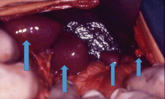
This complex of anomalies is also termed the polysplenia syndrome (polysplenia, absent inferior vena cava, bilobed symmetric liver, malrotation, preduodenal portal vein, bilobed lungs, and cardiac anomalies) and is seen in 10–20 % of patients with biliary atresia (Fig. 42.2) .

Fig. 42.2
An intraoperative photograph showing polysplenia in a patient with biliary atresia
Clinical Features
The initial symptoms of biliary atresia are indistinguishable from neonatal jaundice .
Infants with biliary atresia are typically full term.
The symptoms are seen usually between 1 and 6 weeks of life.
These include:
Jaundice which is prolonged.
Clay-colored stools. The color of the stools may be slightly yellow due to the intestinal epithelial cells, which are shed in the stools. These cells are stained yellow from the generalized jaundice.
Dark urine.
Hepatomegaly may be present early, and the liver is often firm or hard to palpation.
Splenomegaly suggests progressive cirrhosis with portal hypertension .
Differential Diagnosis
The list of causes of neonatal jaundice is long and includes the following :
Alagille syndrome
Biliary hypoplasia
Caroli disease and choledochal cyst
Cystic fibrosis
Cytomegalovirus infection
Galactosemia
Neonatal hemochromatosis
Herpes simplex virus infection
Lipid storage disorders
Rubella
Syphilis
Toxoplasmosis
Investigations
Serum bilirubin (total and direct): Conjugated hyperbilirubinemia, defined as any level exceeding either 2 mg/dL or 20 % of total bilirubin, is always abnormal.
Alkaline phosphatase.
5’ nucleotidase, gamma-glutamyl transpeptidase (GGTP), serum aminotransferases, serum bile acids: A markedly elevated alanine aminotransferase level (> 800 IU/L) is more in favor of neonatal hepatitis.
Stay updated, free articles. Join our Telegram channel

Full access? Get Clinical Tree


