Fig. 9.1
Slate-gray patches affecting the neck (courtesy of Carola Duran McKinster, MD)
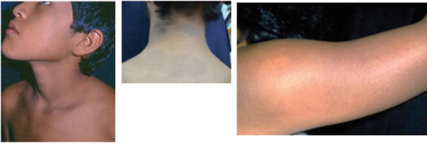
Fig. 9.2
Ashy gray macules and patches affecting the face and neck (courtesy of Carola Duran McKinster, MD)
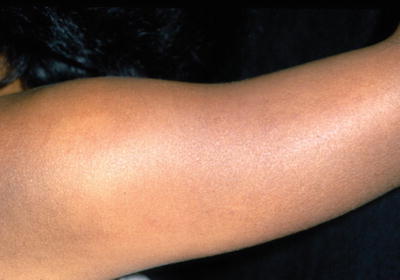
Fig. 9.3
Gray to lead-colored patches affecting the upper arm (courtesy of Carola Duran McKinster, MD)
The etiology of EDP remains unknown although several associations have been noted including benzodiazepines [14, 15], thyroid disease [16, 17], environmental allergens such as the pesticide chlorothalonil [18], toxins such as ammonium nitrate [19], and the fungicide fusilade [17].
Histopathology of the inflammatory border may demonstrate lichenoid dermatitis with vacuolization and necrosis of basal cells and occasional colloid bodies. The papillary dermis is edematous with incontinence of the pigment and shows a mild-to-moderate inflammatory lymphohistiocytic infiltrate intermingled with melanophages [20].
EDP can be differentiated from fixed drug eruption from its gray color, lack of underlying drug trigger, absence of eosinophilia, and less extensive basal layer hydropic degeneration.
In argyria, the mucous membrane and sun-exposed areas are affected and histology shows distinct gray-brown granules in macrophages and adnexal structures.
Prognosis
No treatment has been consistently effectively for EDP
Childhood EDP likely to have significant improvement
Sun protection is advised
Although EDP is unlikely to resolve in adults [1, 10, 13–16, 19, 21–29], an eventual improvement or resolution of the lesions is more often observed in children. The majority of prepubertal patients will be expected to experience significant improvement after 2–3 years [2, 9] and recurrences have never been reported after clearance. No treatment has been consistently effective for EDP and it is recommended to use sun protection to avoid lesional prominence until the lesions clear. Topical steroids and hydroquinones are not useful. Recently, dapsone and clofazimine have been reported to prevent disease progression in adults [12, 17, 30]. Other anecdotal reports have used oral corticosteroids, tetracyclines, ultraviolet light, isoniazid, and griseofulvin.
Summary
Unlike adult EDP, childhood EDP is less common in non-white populations. It tends to affect the truck and proximal extremities. The etiology is not well understood and no therapy has been used successfully to treat EDP. However, in childhood EDP, there is a good chance of complete clearance with time.
References
1.
Ramirez O, Lopez Lino DG. Current status of ashy dermatosis. Synonym-erythema dyschromicum perstans. Med Cutan Ibero Lat Am. 1984;12(1):11–8.PubMed
2.
3.
Epps RE. Case reports: selected dermatoses in children of color. J Drugs Dermatol. 2007;6(1):78–82.PubMed
4.
Imanishi H, Tsuruta D, Kobayashi H, Ishii M, Nakagawa K. Two cases of unilateral ashy dermatosis. Case Rep Dermatol. 2011;3(1):1–4.CrossRefPubMedCentralPubMed
5.
Keisham C, Sarkar R, Garg VK, Chugh S. Ashy dermatosis in an 8-year-old Indian child. Indian Dermatol Online J. 2013;4(1):30–2.CrossRefPubMedCentralPubMed
Stay updated, free articles. Join our Telegram channel

Full access? Get Clinical Tree


