13.3 Arthritis and connective tissue disorders
Box 13.3.1 lists some of the important forms of chronic arthritis and connective tissue disorders of childhood.
Arthritis in children
Arthritis is inflammation of, or related to, a joint and symptoms of arthritis may include:
In the evaluation of the acute onset of joint inflammation, infection or trauma must be considered. Infection may occur as primary septic arthritis or extension of infection from a nearby focus of osteomyelitis into the joint space (see Chapter 12.2). Meningococcal septicaemia and Haemophilus influenzae type b meningitis may be complicated by septic arthritis, or by sterile reactive arthritis. Where trauma is suspected with joint swelling, both accidental and non-accidental injury must always be considered. Inflammation in a joint may also result from bleeding into the joint in haemophilia (see Chapter 16.2), from chloroma associated with neoplastic disease, or rarely from foreign-body penetration.
Juvenile idiopathic arthritis
In 1996, efforts to harmonize research and clinical care in juvenile forms of chronic arthritis resulted in the development of an international classification system, which is now widely recognized. The most recent revision of this classification is detailed in Box 13.3.1. The three main broad clinical presentations are:
Oligoarthritis
Oligoarthritis, defined as four or fewer joints involved during the first 6 months after onset of symptoms, is the commonest form of JIA, accounting for over 60% of cases. Most children present between 1 and 4 years of age, girls twice as frequently as boys. Knees are affected most commonly, followed by ankle (and subtalar) joints, wrists and elbows. Hip disease is rare, and a child presenting with isolated hip involvement must be investigated carefully for disorders such as septic arthritis or osteomyelitis and, in the appropriate age groups, avascular necrosis of the femoral head (Perthes disease) and slipped femoral capital epiphysis (see Chapter 8.1).
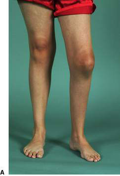
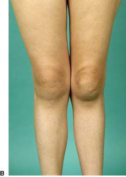
There is usually no systemic disturbance in oligoarthritis. Radiographs may show soft tissue swelling, effusions and widening of the joint space, but erosions are rare. Where treatment has been delayed, epiphyseal overgrowth and lengthening of the involved limb is common. Untreated children ultimately develop erosive disease, whereas growth plate involvement may result in arrest of linear growth and limb deformity (Fig. 13.3.1A,B). More than 70% have a positive serological test for antinuclear antibody (ANA), and rheumatoid factor is usually negative. Oligoarthritis onset has the greatest association with anterior uveitis (see below), which if untreated can cause blindness. Approximately 25% of patients with oligoarthritis will develop progressive polyarthritis after the first 6 months, and are classified as having extended oligoarthritis.

Polyarthritis
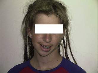
Asymmetrical large and small joint involvement is usual. Hip disease may occur, particularly later. Untreated polyarthritis may cause severe disability, joint deformities, asymmetrical overgrowth (particularly knees) and undergrowth (temporomandibular joint and mandible; Fig. 13.3.2), destruction, ankylosis (especially the wrist and cervical spine) and considerable muscle wasting.
Systemic arthritis
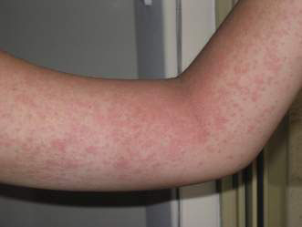
Systemic arthritis affects both sexes equally and accounts for about 10% of cases of JIA. The initial presentation is often with daily or twice-daily spiking fevers, usually above 39 °C, returning to normal between spikes. Affected children are very irritable and movement appears painful, although joints may not appear inflamed. A classical evanescent rash, usually salmon-pink macules, may occur on the upper trunk, arms and thighs associated with fever, a warm bath or scratching the skin (Koebner phenomenon) (Fig. 13.3.3). Notably, in some individuals, particularly darker-skinned children, the rash may be more urticarial and papular.

Other forms of juvenile idiopathic arthritis
Eye disease in juvenile arthritis
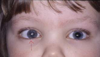
Inflammatory disease of the uveal tract (uveitis or iridocyclitis) can complicate most forms of JIA, and is an important cause of acquired paediatric eye disease and blindness in developed countries. Young girls with oligoarthritis who are ANA-positive are at highest risk, whereas young boys may be more refractory to treatment (Fig. 13.3.4).
A granulomatous pan-uveitis or posterior uveitis may occur in paediatric sarcoidosis, which is rare.
Investigation of chronic arthritis in childhood
Pathology and laboratory findings
Useful initial investigations are listed in Box 13.3.2. Common findings on investigation in a child with one of the arthritis or connective tissue disorders are:
• hypochromic microcytic anaemia
• raised erythrocyte sedimentation rate (ESR) and C-reactive protein (CRP) level
• autoantibodies, for example antinuclear antibody (ANA) and, less commonly, rheumatoid factor (RF+ polyarthritis) or anti-cyclic citrullinated peptide (anti-CCP) antibodies
• inflammatory synovial fluid (white cell count > 2000/mm3, and protein)
• histological evidence of chronic inflammation in synovium.
Box 13.3.2 Initial investigations in suspected juvenile arthritis
• Full blood count (FBC) and film
• Erythrocyte sedimentation rate and/or C-reactive protein
• HLA-B27 (children aged ≥ 6 years, especially males)
• Liver function, coagulation profile, iron studies and renal function tests in those who are systemically unwell
• Radiography of major involved joints (may X-ray contralateral joints for comparison)
Stay updated, free articles. Join our Telegram channel

Full access? Get Clinical Tree


