Fig. 13.1
(a) A young woman with proximal femoral deficiency. Her management was with Symes amputation and knee fusion. She is celebrating her first Paralympic gold medal in three-track skiing at Salt Lake City. (b) The same woman as she achieves another Paralympic medal, this time in track cycling. Overall she has now medaled in four Paralympic games
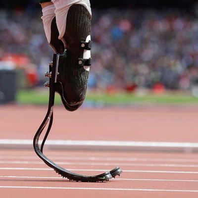
Fig. 13.2
Cheetah type carbon blades. These enable highly competitive performance in a number of sports including track and field, basketball, and football
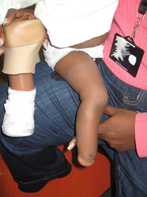
Initial enthusiasm for limb lengthening reconstruction for congenital deformities was very high. As the difficulties and complications of such interventions came to light, especially those encountered with “heroic” lengthenings, a reconsideration of the role of amputation has been appropriate [1–7]. Let us consider the example of a child born with fibular hemimelia, or congenital absence of the fibula, and we will compare Syme amputation to correction with the Ilizarov method. A child with a fibular hemimelia with a well-formed, four ray foot and a 10 % limb length discrepancy can expect an excellent result with one tibial lengthening and a contralateral epiphysiodesis and is not usually a candidate for amputation (Fig. 13.3). At the other end of the spectrum, a child with a very short tibia, marked anterolateral bowing, and a two ray foot can expect to achieve his or her full athletic potential with a single surgery and appropriate prosthetic management following amputation done at 11 months of age. (Fig. 13.4a, b) An Ilizarov approach would require three or more periods of frame management, with considerable loss of childhood experiences, with an end result of compromised function and cosmesis. The decision for management of the child with a deformity in between these two extremes is more difficult. The decisions are complex and emotionally laden and require extensive knowledge of all aspects of proposed treatment, and consideration of family and social dynamics [8, 9].
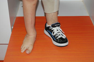
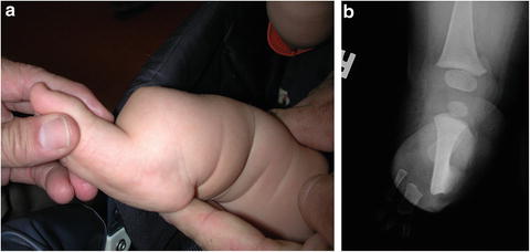

Fig. 13.3
A boy’s foot with fibular hemimelia. This four-rayed foot was functional and had good mobility. Note the Symes prosthesis on the other side, which had a more severe fibular deficiency

Fig. 13.4
(a) A more severe fibular hemimelia with marked shortening and angulation of the lower limb. The two-rayed foot was subsequently converted to a Symes amputation. (b) A radiograph of the same leg showing marked shortening and angulation of the tibia
Treatment Concepts
Congenital deficiencies involving the lower extremities present with varying degrees of severity, some of which are best treated with amputation. Amputation in this context is often one part of a complex strategy involving removal of some bony and soft tissue elements while reconstructing other anatomic components. When applied appropriately, amputation becomes a very positive step in achieving the best functional and cosmetic outcome for the child (Box 13.1). Conditions in which amputation is often useful include congenital femoral deficiency, fibular hemimelia, and tibial hemimelia. Amputation for congenital pseudarthrosis of the tibia may be appropriate after failure of other methods to obtain a stable union with a functional extremity, and occasionally amputation is chosen as the primary management for this condition.
Deformity and dysfunction following severe trauma may also be an indication for amputation, either primarily or following attempts at reconstruction. Primary amputation may be indicated in young children when major epiphyses are injured in such a way that future growth is severely limited.
Many methods of limb reconstruction are appropriately used maintain lower extremity function after tumor excision. Allograft and internal prosthetic reconstruction have made a positive impact on the management of malignant and aggressive benign tumors in children. Amputation at times becomes a solution to intractable complications such as infection and implant failure after these initial efforts. Amputation remains the best primary treatment option when the size and location of the tumor exceeds the limits of reconstructive options [10–18]. In addition, amputation may be appropriate when the patient presents with wide-spread disease, and amputation can provide pain relief and the ability to ambulate over a shortened life span.
Box 13.1.
Amputation as a positive event:
PFFD
Fibular hemimelia
Tibial hemimelia
Trauma
Others
Patient and Family Management
Management options for these abnormalities also vary greatly depending not only on the severity of the condition, but also on the available medical expertise and prosthetic support. Early on, the parents should be introduced to and educated about treatment methods which involve amputation as well as those which involve other methods of complex reconstruction. This education is markedly enhanced when such parents trying to make a decision for their child meet other parents, some of whose children are being managed by amputation and others by limb lengthening reconstruction. These encounters allow parents to ask other parents questions which they would not ask their doctors, and in fact, which the doctors are not really very good at answering.
Historically, amputation was a last resort, dreaded by the surgeon and feared by the patient and caretakers. As prosthetic devices have become more functional and cosmetic, and sports prostheses have allowed people with amputations to compete in sports at very high levels, the concept of amputation as a positive strategy has taken hold in society. Parents and children need to be informed about modern amputation techniques and prosthetic fitting which provide high function with a minimum number of surgical encounters and morbidity (Box 13.2). They will need the knowledge with which to compare amputation to limb lengthening reconstruction, typically requiring multiple procedures over time. They should be provided with honest information about complications and outcomes of all methods, frequency and duration of hospitalizations, and the availability and quality of prosthetic care.
Box 13.2
Education is essential:
By physician
By other parents (who have had a child with a similar diagnosis)
With psychologic support
The comparative costs of prosthetic management and complex reconstruction are difficult to assess. Several studies have shown conflicting conclusions and consideration of all relevant factors is difficult. A most important cost to consider is the lost childhood or adolescence which occurs with multiple episodes of frame application and hospitalizations to manage complications [19–21].
Specific Conditions
Congenital Femoral Deficiency
There is great variation of femoral anatomy among children with congenital femoral deficiency (CFD), and several classifications are useful. The Gillespie classification [22–24] is based on length of the femur at presentation (Fig. 13.5). In type A the femur is at least 50 % as long as the normal femur, and in type B it is less than 50 % of the normal length. In type C there is almost no development of the proximal femur. The classification relates in general to treatment recommendations. The Hamanishi classification [25] illustrates the wide variety of anatomic presentations, but is not specifically tied to treatment (Fig. 13.6). The Paley classification [26] adds consideration of the mobility of the femoral head within the acetabulum, and is useful for some reconstruction methods (Fig. 13.7).
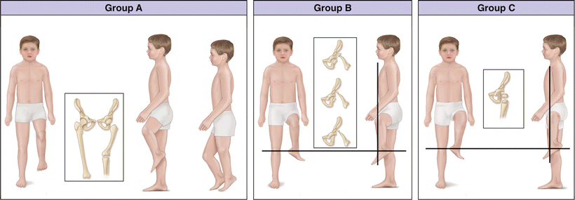
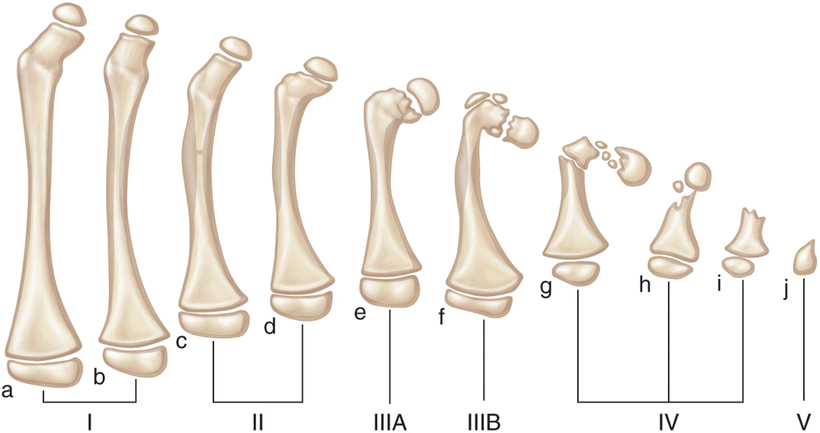
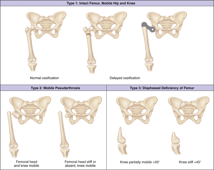

Fig. 13.5
The Gillespie classification of femoral deficiency. In type A the femoral length is more than 50 % of the normal side. In type B, the length of the femur is 50 % or less than the other femur. In type C, there is almost no femoral length. This image was published in Tachdjian’s Pediatric Orthopedics, Herring JA, Limb deficiencies, 965–974, Copyright Elsevier 2014

Fig. 13.6
The Hamanishi classification of femoral deficiency. This classification notes the multiple variations of the deformity. This image was published in Tachdjian’s Pediatric Orthopedics, Herring JA, Limb deficiencies, 965–974, Copyright Elsevier 2014

Fig. 13.7
The Paley classification of femoral deficiency. This classification takes note of hip and knee mobility. This image was published in Tachdjian’s Pediatric Orthopedics, Herring JA, Limb deficiencies, 965–974, Copyright Elsevier 2014
Treatment in Gillespie Type A
These patients, with at least 50 % of normal femoral length can usually be managed with lengthening and reconstructive methods. The outcome is best when the hip is stable and well formed, and when the upper femoral deformity or deficiency can be corrected. The greater the shortening of the femur, the greater is the number of corrective and lengthening procedures that will be required. The lengthening process in this condition is difficult and a number of authors report a high frequency of complications and failures because of shortening and contracture of all the tissues in the thigh [27–29].
Gillespie Types B and C
In these patients the femur is less than 50 % of the normal length, and lengthening and reconstruction will generally require three or more episodes of lengthening. Each of these will consume months of time, and complications often increase with successive lengthening. Likewise, the ultimate function likely decreases as the underlying anatomy is more deficient in cases with substantial femoral shortening. Thus, in this situation some variation of amputation is usually appropriate (Box 13.3). Patients with bilateral femoral deficiency are almost never candidates for amputation, nor are those with major upper limb anomalies requiring the feet for manual activities (Fig. 13.8).
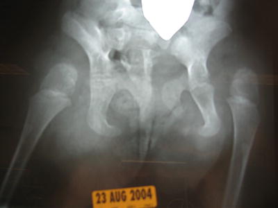

Fig. 13.8
A radiograph of severe bilateral femoral deficiency, Gillespie type C, in which there is no femoral formation and the tibias articulate with the pelvis. Treatment alternatives are minimal in these children. This image was published in Tachdjian’s Pediatric Orthopedics, Herring JA, Limb deficiencies, 965–974, Copyright Elsevier 2014
The treatment options in CFD can best be understood relative to the presenting anatomy. Factors to consider include (1) the anatomy of the hip and its musculature, (2) relative shortening of the femur, (3) the range of motion of the knee, (4) the anatomy of the foot. The anatomy of the hip varies from relatively normal to severe deformity or sometimes absence of the femoral head and acetabulum. When possible, the anatomy of the upper femur should be reconstructed to restore bony continuity of the femur and restore the femoral neck shaft angle. Also, any acetabular dysplasia should also be corrected (Fig. 13.9). When the femoral head has not developed, or is immobile in the acetabulum, hip reconstruction is generally not feasible.
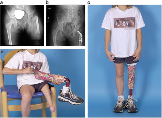

Fig. 13.9
(a) A Gillespie type B deficiency with less than 50 % of femoral length. There is a short femoral neck with a marked varus deformity. (b) A radiograph of the pelvis after proximal femoral valgus osteotomy and acetabuloplasty. (c) The same patient with her prosthesis. Her anatomic knee is intact and flexes just at the top of the prosthesis. She is an active varsity cheerleader with no functional limitations. (d) Her shortened thigh segment is evident as she sits without her prosthesis
Knee arthrodesis with Symes amputation has been used for many years and provides good function. The child can be fitted with a prosthesis at walking age incorporating the foot in the prosthetic socket, or a Symes amputation can be done at walking age to facilitate prosthetic fitting. Arthrodesis of the knee is usually done at an older age, often age 3 or 4 years, when there is significant ossification of the knee structures. The arthrodesis improves the gait by reducing the flexed position of the thigh segment and aligning the mechanical axis of the limb under the weight line of the body. To have appropriate length of the thigh segment after arthrodesis, the upper tibial epiphysis is usually fused to the distal femoral metaphysis with removal of the distal femoral epiphysis (Fig. 13.10). Ideally the end of the thigh segment should be at least 10 cm shorter than the contralateral knee to allow room for the prosthetic knee components. The relative growth of this segment is less than the normal femur by the amount of growth of the contralateral distal femoral epiphysis, since the homolateral epiphysis was removed at the time of knee fusion. As a practical matter, if the child is reaching adolescence and the knees are at the same level with a shorter prosthetic tibial segment, the surgeon should consider performing a proximal tibial epiphysis on the side of the limb deficiency.
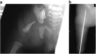

Fig. 13.10
(a) A radiograph of a type B femoral deficiency. Note the absence of femoral head or acetabular development. (b) The same patient after arthrodesis of the knee and Symes amputation
As an alternative, rotation of the distal limb to achieve a Van Nes effect may be done at the time of knee arthrodesis [30, 31]. When there is no hip stability, the distal segment has a tendency to derotate with growth, and may have to be repeated.
When the proximal hip anatomy is not amenable to reconstruction, several procedures have been developed in an attempt to reduce the abductor limp which is usually quite noticeable. The most successful has been that developed by Ken Brown in which the femur is rotated 180° and fused to the pelvis in a vertical position [30, 31] (Fig. 13.11). In this position flexion of the rotated knee becomes hip flexion. The fusion of the femur to the pelvis, with full rotation turns the foot backwards with the toes facing posteriorly. The foot is placed in equinus into a prosthetic socket so that the ankle controls the prosthetic knee. When the ankle dorsiflexes the prosthetic knee flexes and when the foot plantar flexes the knee extends. The anatomic ankle also provides proprioception of “knee” position which is an important advantage for the patient, for example when descending stairs.
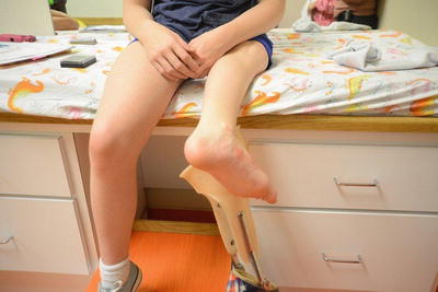

Fig. 13.11
A 10-year-old girl with a Van Nes type rotationplasty after tumor resection
Box 13.3.
Options with amputation:
Femoral length <50 %
Hip reconstruction
Knee fusion
Femoral-pelvic fusion
Symes amputation
Rotationplasty
Congenital Fibular Deficiency
While several classifications of congenital fibular deficiency (or fibular hemimelia) are used, we prefer the Birch classification [32], which is based largely on the severity of foot deformity and the relative limb length (Table 13.1). In general, a patient whose foot can be made plantigrade and which has three or more rays may be a candidate for lengthening. When the overall limb length inequality is 10 % or less, lengthening is preferred. With discrepancies between 10 and 30 %, either method may be appropriate. With discrepancies greater than 30 %, often requiring more than two limb lengthening procedures, we usually recommend an amputation (Box 13.4) (see Figs. 13.4 and 13.12).
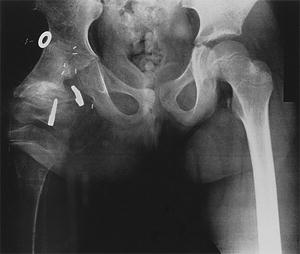
Table 13.1
Proposed classification of congenital fibular deficiency based on clinical deformity and treatment based on classificationa
Type | Characteristic | Treatment anticipated |
|---|---|---|
Type 1 (foot preservable) | ||
1A | <6 % inequality | No treatment or orthosis or epiphysiodesis |
1B | 6–10 % inequality | Epiphysiodesis ± lengthening |
1C | 11–30 % inequality | 1 or 2 lengthenings ± epiphysiodesis or extension orthosis |
1D | >30 % inequality | >2 lengthenings or amputation or extension orthosis |
Type 2 (foot nonpreservable) | ||
2A | Functional upper extremity | Early amputation |
2B | Nonfunctional upper extremity | Consider salvage |

Fig. 13.12
A radiograph of a patient who had a Brown procedure. The proximal femur with a tumor was removed and the distal femur was rotated 180° and fused to the pelvis in an extended position. The staples are placed to stop the growth of the distal femoral epiphysis
Our preferred amputation technique for fibular hemimelia is a variation of the Symes procedure in which the ankle is disarticulated without removing either malleolus [33]. The heel pad remains as an excellent weight bearing structure, and the incision is anterior away from the weight bearing surface (Figs. 13.13 and 13.14). Some surgeons prefer the Boyd modification with fusion of the calcaneus to the distal tibial articular surface.

